
In an angiography suite, a neuroradiologist threads a catheter up through a vein and into the head, finds a stretch where one of the brain's large drainage channels has narrowed, and props it open with a small metal mesh. The patient's problem was pressure, too much of it, inside a skull that cannot expand to accommodate any. For some people with idiopathic intracranial hypertension, the procedure works and the pressure comes down. What has been missing is a satisfying account of why a blood vessel should have so much say over the pressure of the fluid bathing the brain.
A study published in Nature Neuroscience on 22 July proposes an answer, and it runs through a set of vessels that biologists only recently began taking seriously: the lymphatic channels threaded through the membranes wrapping the brain. Combining MRI scans of patients with a mouse experiment, the researchers found that keeping pressure inside the head under control, and clearing fluid out of the brain, depends on those lymphatic vessels. The big veins appear to be where blood flow tells them what to do.
Why it matters: Dangerously high pressure inside the skull is still managed with fairly blunt tools, and the reasons a given treatment helps have been more observed than understood. A mechanism that links venous blood flow to the brain's own drainage gives clinicians something specific to aim at.
The work comes from El Kamouh et al. at the Paris Brain Institute, part of Sorbonne University and INSERM, working with colleagues at Pitie-Salpetriere Hospital in Paris and at Yale School of Medicine. The condition at the centre of it, idiopathic intracranial hypertension, is defined by raised pressure inside the skull together with narrowings, called stenoses, in the dural venous sinuses. Those are the wide venous channels running through the tough membrane that lines the inside of the skull. Stenting the narrowed segment can relieve the pressure, which is a strong hint that venous flow matters, but a hint is not a mechanism.
What the scans showed
The team began with people, imaging patients who had the condition alongside healthy volunteers. In the patients, the venous narrowings came with two things visible on MRI: a changed pattern of fluid around the veins, and swelling in the brain tissue itself. That is a correlation rather than a cause, and the authors treat it as one. But it pointed somewhere specific, at the fluid immediately surrounding the drainage vessels rather than at the vessels alone.
Tying off a vein in a mouse
To turn that association into something testable, the researchers built an animal version of restricted venous outflow by tying off the jugular vein, the main route by which blood leaves the head. The mice responded much as the imaging predicted. Pressure inside the brain rose, though only temporarily, the tissue swelled, and the brain's ability to clear fluid was impaired. The unexpected part was what happened to the lymphatic vessels in the meninges. They came out defective, meaning a blockage on the blood side had damaged the drainage side.
Which part is actually doing the work
Establishing that two things happen together is not the same as showing one depends on the other, so the team removed the meningeal lymphatic vessels and repeated the experiment. Losing them raised pressure inside the brain in ordinary mice as well as in the ones with a tied-off vein, which puts these vessels squarely in the business of pressure regulation. The sharper result came afterwards. Ligated mice with their lymphatics intact eventually recovered their fluid clearance. Ligated mice without them did not. Recovery, in other words, ran through the lymphatic vessels.
The authors put their conclusion plainly. These findings implicate MLVs in the control of intracerebral pressure and establish the dural venous sinuses as critical platforms where venous flow directs MLVs to ensure brain fluid clearance, they write, using the field's shorthand for the meningeal lymphatic vessels. The veins are not just pipes carrying blood away. They are the surface along which the brain's drainage system takes its instructions.
What the study can't say yet
The human side of this is imaging, not treatment. Patients were scanned and compared with controls; nobody was stented and followed to see whether their lymphatic vessels changed, so the study does not demonstrate that stenting works by way of this pathway in people. The mouse work carries the causal weight, and a tied-off jugular vein is a deliberately crude stand-in for the subtler narrowings seen in patients. The pressure rise in the mice was explicitly transient. What remains unresolved is whether the lymphatic vessels in a person with long-standing raised pressure are damaged in the same way, and whether anything can be done about it.
Quick questions
What are meningeal lymphatic vessels? Drainage channels running through the membranes that cover the brain. Long overlooked in anatomy textbooks, they are now an active area of study, and this work places them in the control of pressure inside the skull.
Does this explain why venous stenting helps patients? It offers a plausible route, not a demonstration. Stenting's benefit is established clinically, but this study did not test stented patients.
What is the one-line takeaway? Blood flow through the brain's big veins appears to set the working condition of its lymphatic drainage, which is what keeps pressure in check and fluid moving out.
El Kamouh et al. "Cerebral venous blood flow regulates intracerebral pressure and brain clearance via meningeal lymphatic vessels." Nature Neuroscience, 2026. doi.org/10.1038/s41593-026-02358-1
PubMed PMID: 42487032.
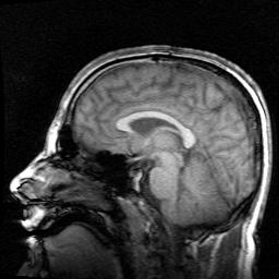
Image: Magnetic resonance image of a human brain, side view. Oliver Stollmann, Attribution (CC BY) license, via Wikimedia Commons.
Comments