
There is a stretch early in life when a young brain is unusually willing to be rewired by whatever it encounters. A kitten that has one eye kept shut during this window can lose vision in that eye for good, even after the eye is reopened, because the visual system reorganizes around what it actually sees. The same principle, in gentler forms, shapes how children learn language, sound, and sight. Then the window narrows. The brain stops rewriting itself so freely and settles into something more fixed. Neuroscientists have long known these "critical periods" exist. What has been harder to explain is what actually pulls the window shut.
A study published in Nature on May 20, 2026 by researchers at Harvard Medical School (Gegenhuber et al.) points to an unexpected participant in that closing: the receptor for the body's main stress hormone, working not in neurons but in the brain's support cells. The finding reframes cortisol-family signaling, usually discussed in the language of stress and fear, as part of the ordinary machinery that ends childhood plasticity.
Why it matters: Critical periods set how easily a young brain can be reshaped by experience, and this work ties their closing to the body's stress-hormone system. That hints that the timing and amount of early-life stress could, in principle, influence brain development and susceptibility to neuropsychiatric disease.
What the researchers found
The team, led by Bruno Gegenhuber in the laboratory of Michael Greenberg, studied the primary visual cortex of mice as it matured after birth. To watch the process cell by cell, they used paired single-cell sequencing, reading both which genes each cell was switching on and which stretches of its DNA were becoming accessible to be read. That combination let them track, across development, how each type of brain cell responded as the animal's eyes opened and light began to arrive.
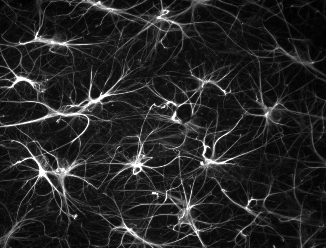
Neurons behaved much as expected, running activity-driven gene programs. The surprise was in the astrocytes, the star-shaped support cells long treated as mere scaffolding. The authors report that "light exposure drives astrocyte maturation," a role for these cells that had gone largely unrecognized. Light exposure drove these astrocytes to mature, and it did so by recruiting the glucocorticoid receptor, the protein encoded by the gene Nr3c1 that normally responds to stress hormones, onto specific regions of the astrocytes' DNA. That receptor then switched on a broad program of genes. As the astrocytes matured under this program, the brain's window of high plasticity closed. In the authors' framing, astrocyte glucocorticoid receptor signaling restricts neuronal plasticity.
In other words, one of the cues that helps end the malleable phase is not coming from the neurons doing the learning. It runs through the surrounding cells, and it uses the same hormonal receptor the body relies on to handle stress.
Why the stress connection matters
That overlap is what gives the work its reach beyond visual development. Glucocorticoids are the hormones that surge during stress. If the receptor for those hormones is embedded in the timetable that closes plasticity, then the amount and timing of stress a young animal experiences could, in principle, nudge that timetable. The authors note that this glucocorticoid control of astrocyte maturation may contribute to the effects of early-life stress across the brain, and that disrupting the process could raise susceptibility to neuropsychiatric disease.
The researchers also report that the gene program driven by the astrocyte glucocorticoid receptor is partially conserved in human brain development. The same regulatory logic appears to be present, in part, in maturing human tissue, which is why the finding is being read as more than a quirk of mouse vision.
What the study can't say yet
This is a mouse study, and most of it centers on one region, the visual cortex. Critical periods differ across brain systems and species, and what governs the visual window need not govern the windows for language or emotion in the same way. The link to human biology rests on the observation that the gene program is partly shared, not on experiments in people.
The connection to early-life stress and neuropsychiatric disease is also a hypothesis the data point toward, not a demonstrated cause. The paper shows that a stress-hormone receptor participates in normal astrocyte maturation and plasticity closure under ordinary light exposure. It does not show that a stressful childhood, in a mouse or a person, reshapes plasticity through this pathway and produces disease. Those are the experiments that would come next: manipulating stress and glucocorticoid signaling during development and tracing the downstream effects on the brain and behavior.
It is also worth being precise about direction. The study describes a mechanism that helps close the window, part of normal maturation, rather than a switch that could be flipped to reopen it. Whether interfering with astrocyte glucocorticoid signaling could extend or reopen a critical period, and whether that would even be desirable, is unknown.
What the work does establish is a genuine shift in where to look. For decades the closing of critical periods was framed largely as a story about neurons and inhibitory circuits maturing. This adds a different character to the account, the astrocyte, listening to light and to a stress hormone at once, and helping decide when a young brain stops being quite so easy to change.
Quick questions
Does this mean stress in childhood damages the brain? Not from this study. It shows that a stress-hormone receptor is part of normal astrocyte maturation under ordinary light; it did not test whether a stressful childhood reshapes plasticity or causes disease. That link is a hypothesis for future work.
Was any of this shown in humans? No. The experiments are in mice, mostly in the visual cortex. The only human connection is that the astrocyte gene program is partially conserved in human brain development.
The one-line takeaway? A stress-hormone receptor working inside the brain's support cells helps pull shut the window when a young brain is easiest to change.
Gegenhuber B et al. "Astrocyte glucocorticoid receptor signalling restricts neuronal plasticity." Nature, 2026. doi.org/10.1038/s41586-026-10512-9
PubMed PMID: 42162428.
Image: Yann Bernardinelli, CC BY-SA 3.0, via Wikimedia Commons.
Comments