When two chromosomes break and rejoin in the wrong order, the seam can fall inside two genes and stitch them into one. The hybrid protein that results is often what drives the cancer. But the more useful property of that seam is quieter: the stretch of sequence spanning the join exists nowhere else in the body. It is not in either parent gene, and not in any healthy cell. It is as close to a tumour-specific barcode as cancer genetics offers.
The difficulty has always been that you cannot easily drug a sequence. Drugs bind proteins, and fusion proteins are largely undruggable; where an inhibitor does exist, resistance tends to follow. A group at the Peter MacCallum Cancer Centre in Melbourne has gone after the sequence instead, using an RNA-cutting CRISPR enzyme called PspCas13b programmed to recognise the breakpoint itself. Because the enzyme finds its target by matching RNA letter for letter, the seam becomes a legitimate address. They silenced nine different fusion transcripts this way, and against the fusion that defines chronic myeloid leukaemia it worked on both the standard version and on the mutant that defeats the existing drugs.
Why it matters: Gene fusions appear in about 20% of human cancers and act as genuine drivers in roughly 16.5% of cases, and More than 3297 unique fusion transcripts have been described across multiple cancer types. Most have no drug. An approach whose targeting rule is simply the patient's own breakpoint sequence is, in principle, as general as sequencing is.
The study, by Hu et al. at the Peter MacCallum Cancer Centre and the University of Melbourne, appears in Science Advances. Every experiment is in cultured cells.
Specificity that comes from the design, not from tuning
Positioning the guide so the breakpoint sits near its centre means neither normal parent transcript is a match. That is specificity by construction rather than by optimisation, and the checks back it up: the enzyme reduced the fusion transcript while leaving untranslocated copies of both parent genes alone, did not chew through a highly expressed unrelated reporter, and when deliberately pointed at a dispensable target produced detectable changes in only two endogenous proteins by mass spectrometry.
The comparison with the existing drug is the sharper point. Imatinib blocks the kinase activity of the fusion protein but leaves the protein itself in place at normal levels. Destroying the message removes the protein outright. That distinction matters because fusion proteins also work as scaffolds, holding other molecules together, and shutting off catalysis alone is sometimes not enough to stop them.
Resistance follows the same logic. The T315I mutation that defeats several kinase inhibitors sits in the kinase domain and leaves the breakpoint untouched, so the guide still matches. In drug-resistant cells, silencing pushed them into erythroid differentiation and apoptosis.
A measurement trap the paper walks into and then explains
The most useful thing here is a result that initially looked wrong. Short-read RNA-seq analysis showed no reduction in overall BCR::ABL1 mRNA levels, and the authors note that This observation was unexpected given the efficient knockdown of BCR::ABL1 protein. The protein was gone; the message, measured in bulk, was not.
The explanation is that the enzyme nicks rather than demolishes. Only a region of roughly 500 bases around the cut site is degraded, and the authors conclude that Our data suggest that Cas13-mediated target cleavage readily degrades regions proximal to its binding site, whereas distal regions are relatively stabilized. Long-read sequencing showed that the transcripts which survive carry scrambled junctions, frequently out of frame and incapable of being translated, which reconciles the two observations: the RNA is still countable but no longer readable.
The practical consequence reaches well beyond this paper. If cleavage is local, then a standard RT-qPCR measurement of knockdown depends almost entirely on where the primers happen to sit, and the same behaviour has been reported for a different Cas13 variant. The authors' recommendation is to place primers flanking the cut site. Any published Cas13 knockdown figure measured otherwise is, on this evidence, difficult to interpret.
What the study can't say yet
No animal was treated. The entire body of work is cell lines, including patient-derived leukaemia and sarcoma lines, and the authors describe it as a proof-of-concept study. Nothing here shows a tumour shrinking in a living organism.
Delivery, which is the actual obstacle to this becoming a therapy, is barely engaged. The leukaemia line used resists ordinary transfection, so the enzyme had to be introduced by nucleofection or by lipid nanoparticles in a dish. Getting an RNA-guided enzyme into cancer cells distributed through a patient's marrow and blood is a different problem of a different size.
Two further caveats sit inside the mechanism. RNA targeting is inherently temporary, which is a safety advantage and a durability problem, and no experiment here measures how long silencing persists or what repeat dosing does. And the religation finding cuts both ways: the authors report that A fraction of target RNA molecules displayed internal deletions, suggestive of putative RNA ligation upon cleavage, with aberrant transcripts reaching about 50% in one cell line and less in others. They read those products as non-functional, but a repair process whose frequency varies by cell type and whose mechanism is unresolved is not something to assume is harmless.
Quick questions
Could this treat leukaemia? Not yet, and not close. It is a targeting strategy demonstrated in cultured cells, with the delivery question untouched and no animal data at all.
Why go after RNA rather than editing the DNA? Cutting DNA makes a permanent change and risks editing the genome of healthy cells. RNA targeting wears off, which limits the damage a mistake can do, and the breakpoint is just as readable in the message as in the gene.
What is the one-line takeaway? Programming an RNA-cutting CRISPR enzyme to recognise the seam where two genes were fused silenced nine cancer fusions including a drug-resistant leukaemia driver, with the bonus finding that these enzymes cut locally rather than destroying the transcript, which means a lot of published knockdown measurements depend on where the primers were placed.
Hu et al. "Programmable and selective silencing of oncogenic fusion transcripts by CRISPR-Cas13b via breakpoint sequence recognition." Science Advances, 2026. doi.org/10.1126/sciadv.ady0296
PubMed PMID: 42886793. Full text: PMC13674509.
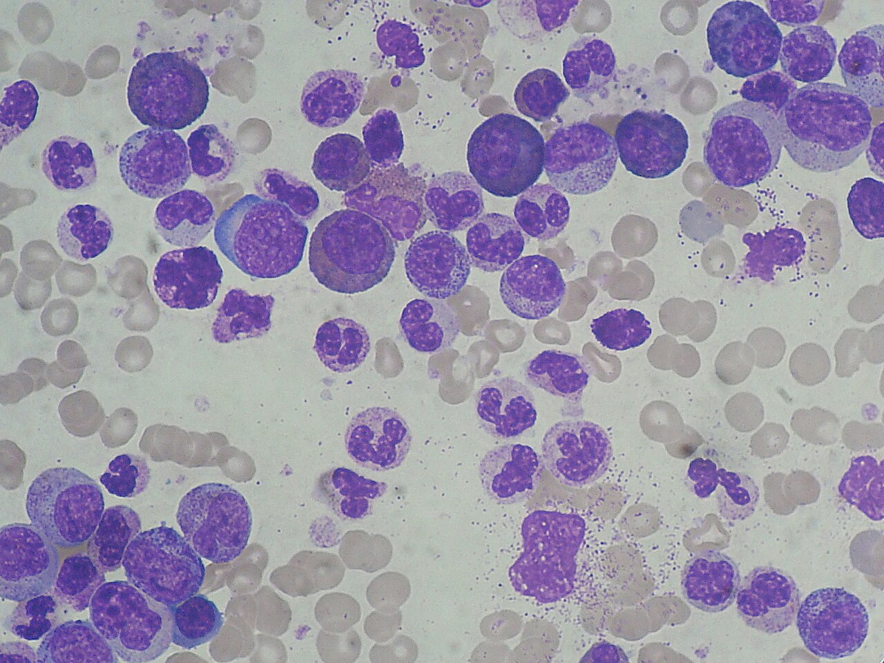
Image: chronic myeloid leukaemia blood smear. Paulo Henrique Orlandi Mourao, CC BY-SA 3.0, via Wikimedia Commons.
Comments