The difficulty with fixing a broken ion channel is that one mutation often breaks it in two incompatible directions. The cardiac sodium channel, NaV1.5, opens to let sodium rush into heart muscle cells and start each beat, then shuts. Some mutations stop it shutting properly, leaving a small current leaking through and stretching each beat out; that is long QT syndrome type 3. Other mutations mean fewer working channels reach the cell surface at all, weakening the initial surge; that is Brugada syndrome. And a third group of mutations does both at once.
That third group is the problem. The drugs available block the leak, but blocking does nothing about the missing channels, and can reduce the initial surge further. Push the other way, sending more channels to the surface, and you deliver more faulty channels, making the leak worse. A group at Columbia University has now built a single protein designed to do both jobs in opposite directions: a nanobody that latches onto the channel and carries two working parts, one that restores proper shutting and one that keeps the channel at the cell surface.
Why it matters: Roughly 15% of marketed drugs target ion channels, but almost all of them work by blocking. A molecule that can add function in one respect while subtracting it in another is a different kind of tool, and the same logic would apply to sodium-channel mutations behind some epilepsies and muscle disorders.
The study, by Fossier et al. at Columbia University, with a co-author at Merck Research Laboratory, appears in Science Advances. The work runs through cultured cells, mouse heart muscle cells and heart cells grown from human stem cells.
Two tools on one carrier
The carrier is a nanobody, a small antibody fragment that binds the channel tightly. Onto it the team attached two things. The first is a short peptide called FixR, derived from a growth-factor-related protein the body already uses to regulate these channels; it encourages the channel to shut properly, suppressing the leak. The second is the business end of an enzyme called Otud1, which strips off a particular type of ubiquitin tag.
That second part is the more interesting half. Ubiquitin tags are the cell's labels for trafficking and disposal, and the specific linkage Otud1 removes is one that marks channels for removal from the surface. Pulling those tags off keeps more channels where they are useful. The specificity turns out to matter: of three enzymes tried on the normal channel, the Otud1 version raised currents, a different one that cuts another linkage type lowered them, and a third that cuts both did almost nothing. On one disease variant, all three raised the current, which the authors read as that variant carrying more of the tags to begin with.
Combined, the construct produced roughly a fourfold increase in current in the strongest case. In mice given an early-stage heart-failure diet, where the surge current had dropped by about half, delivering the construct by virus restored channel levels at the surface and brought the current back.
Why blocking alone was never going to be enough
The existing answer to the leak is a blocker that plugs the channel's pore. Several exist, and a peptide approach from the same lab has already been shown to cut the leak and reduce arrhythmia in rabbits and mice. The limitation is structural rather than a matter of potency: a molecule that obstructs the pore cannot also open the route for more channels to reach the surface, and in some cases blockers reduce the initial surge, which is the opposite of what a Brugada-type mutation needs.
Attempts to run the other way have their own problem. Overexpressing a chaperone protein does push more channels to the membrane, but that protein has other binding partners, so the effect is not confined to the target. For a related channel in the brain, antisense oligonucleotides raise channel levels by correcting a splicing error, but no equivalent handle exists here. The case for putting both functions on one carrier is that neither direction is safe on its own when a mutation causes both faults.
What the study can't say yet
The authors are unusually direct about the gap, and it is a wide one. Nothing here shows the construct prevents an abnormal rhythm. They did not measure arrhythmia susceptibility, conduction speed or calcium handling in a living animal, and they say plainly that whether the molecular changes are also sufficient to restore normal electrical rhythm remains unknown. Restoring a current in a cell is a molecular result, not a clinical one.
The headline claim is also only half-demonstrated in an animal. The whole point is correcting both faults simultaneously, but the mouse model used was an early-stage one that showed only the weakened surge, with no leak to fix. So in a living animal the construct was shown to do one of its two jobs. Proving both at once would need a mouse carrying a mutation that causes both, which the authors name as necessary future work.
Scale is worth noting too. The cell experiments rest on single-digit numbers of cells, typically six to twelve per condition, from two or three separate preparations. That is normal for this kind of electrical recording but it is a long way from a result that has been stress-tested.
Delivery is unresolved. The construct reached mouse hearts inside an adeno-associated virus, which the authors flag as having translational limits, and they float lipid nanoparticles as an alternative. One unexplained observation sits in the data: mice given the construct gained more weight than controls, which the authors attribute tentatively to effects of the virus outside the heart.
Quick questions
Is this close to a treatment? No. It is described by its authors as a proof of concept, and the step it has not taken is showing any effect on heart rhythm in a living animal.
Why use an antibody fragment rather than a drug? Because the two jobs need to happen at one specific protein. The nanobody is the address label, holding both tools against NaV1.5 rather than letting them act across the cell.
What is the one-line takeaway? A single engineered protein corrected both a gating fault and a trafficking fault in a cardiac sodium channel in cells, which is a genuine first, but whether that translates into a steadier heartbeat is untested.
Fossier et al. "A bifunctional actuator to correct loss-of-function deficits in cardiac voltage-gated sodium channels." Science Advances, 2026. doi.org/10.1126/sciadv.aeb2110
PubMed PMID: 42853924.
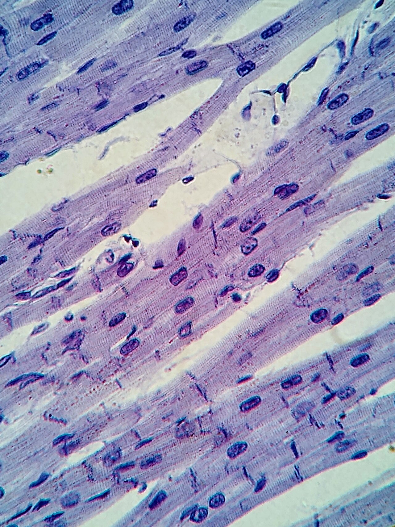
Image: cardiac muscle at 400x magnification. Alexander G. Cheroske, CC BY-SA 4.0, via Wikimedia Commons.
Comments