The constraint on Chlamydia trachomatis is that it cannot afford to build much. Its genome runs to roughly 900 genes, missing whole metabolic pathways, so it lives inside human cells and steals what it needs: amino acids, nucleotides, lipids. That thrift shapes its life cycle. The form that travels between cells, the elementary body, is only 200 to 300 nanometres across and metabolically shut down, its DNA crushed into a dense ball called a nucleoid. Once inside a new cell it converts into a larger, active, replicating form, and the first thing that has to happen is that the ball unpacks.
How it unpacks has been largely unknown. A group at the University of Würzburg, using an imaging trick that physically swells a specimen to beat the resolution limit of light, has now found something unexpected sitting inside that packed DNA: derivatives of sphingomyelin, a lipid the bacterium takes from its host. Around 90% of the nucleoid area carried the lipid label. And the lipid leaves the nucleoid before the DNA decondenses, within an hour of the bacterium entering a cell, making its departure the earliest visible step in the transition.
Why it matters: Chlamydia is the most common bacterial sexually transmitted infection worldwide, and the switch between its dormant and replicating forms is the point at which an infection establishes itself. A lipid involved in throwing that switch would be a target that the bacterium cannot easily mutate away, because it does not make the lipid in the first place.
The study, by Rühling et al. at Julius-Maximilians-University Würzburg, with collaborators at the University of Zürich, Freie Universität Berlin and the MRC Laboratory of Molecular Biology, appears in Nature Communications. The work is done in infected cultured human cells.
Seeing a lipid inside DNA
The method matters here, because the claim depends on it. Conventional light microscopy cannot resolve anything finer than about 250 nanometres, which is the size of the whole bacterium. Expansion microscopy gets around this by embedding the fixed sample in a gel that swells in water, pulling the labelled molecules physically apart so an ordinary confocal microscope can separate them. At eightfold expansion the effective resolution is around 30 nanometres, enough to see structure inside a particle that is otherwise a single dot.
Lipids are awkward to image this way because most of them are not anchored into the gel, so the team used synthetic sphingomyelin derivatives built to be both anchorable and trackable. Alongside this they ran electron tomography of frozen samples, a FRET-based readout that reports whether the probe has been chemically converted, and bulk lipid measurement. The convergence is the argument: the imaging says where, the FRET says the probe is being metabolised rather than merely sticking, and the lipidomics says which species dominates.
An architecture with a front and a back
The elementary body turns out to be organised, not a uniform package. Scoring 105 of them, the injection apparatus the bacterium uses to manipulate host cells sat at one pole, while stacks of internal membrane sat at the opposite pole in 71% of cells. In tomograms where both the nucleoid and those membrane stacks could be identified, the nucleoid was fully aligned with the membranes in 69% of cases and offset but still touching in a further 23%. The genome is not floating free; it is parked against a membrane.
Following infection over time, particles grew from about 200 nanometres to 750 within the first six hours as they converted to the replicating form, and the imaging resolved several morphologically distinct intermediate states along the way, distinguishable by how the DNA inside was arranged. Those intermediates had been postulated but poorly defined. Separately, replicating bacteria held artificially in an arrested state failed to accumulate sphingomyelin, which is the observation that ties the lipid to the developmental stage rather than to the cell generally.
What the study can't say yet
The most important limitation is the probe, and the authors put it plainly. The synthetic lipids carry six-carbon tails, whereas natural lipids typically carry fourteen or more, and chain length changes how a lipid behaves, including how readily it hops between membranes. Thus, TFSM transport and localization could differ from natural long chain SM species, they write, adding that their method cannot currently visualise natural lipids at all. So this is a map of where a short-tailed analogue goes.
The second is that nothing here is causal. The lipid is present, and it leaves before the DNA unpacks, but whether it does anything is open: it remains elusive whether, and if so, how lipids can actively contribute to DNA condensation in C. trachomatis. The energy-saving logic the authors sketch, that hijacking a host lipid is cheaper than synthesising DNA-packing proteins, is a hypothesis, made more interesting by a recent report that chlamydial nucleoid formation does not require those proteins.
Two technical caveats cut the same way. A second lipid probe designed to stain membranes gave a different and more variable pattern, which the authors attribute to the two dyes labelling different lipid pools rather than to one being wrong, but it is an unresolved discrepancy. And in the tomography the nucleoids were not labelled directly; they were inferred from regions where ribosomes were absent. The resolution is also not quite sufficient to say whether the membrane merely abuts the DNA or penetrates it.
Quick questions
Does this suggest a treatment? Not directly. It identifies a host-supplied molecule associated with a critical switch, which is the kind of dependency worth probing, but no drug or intervention was tested.
Why would a bacterium use fat to fold DNA? Possibly economy. Most bacteria use proteins; this one is missing large parts of its metabolism and already imports host lipids in bulk, so using them structurally would avoid building something new.
What is the one-line takeaway? A host lipid was imaged inside the condensed genome of Chlamydia's infectious form and seen leaving just before the genome unpacks, which is suggestive of a role in the switch but, on short-tailed synthetic probes alone, not yet evidence of one.
Rühling et al. "Sphingolipids associate with the chlamydial nucleoid and mark developmental transitions in Chlamydia trachomatis." Nature Communications, 2026. doi.org/10.1038/s41467-026-77974-3
PubMed PMID: 42816552.
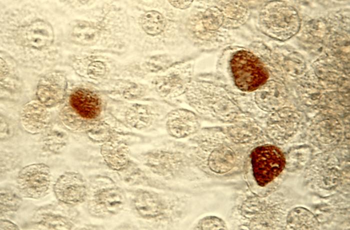
Image: Chlamydia trachomatis inclusion bodies in cultured cells. Marcus007, public domain, via Wikimedia Commons.
Comments