
Schistosomes are the exception among parasitic flatworms. Most are hermaphrodites; these have separate sexes, chromosomally determined, and yet males and females look identical through almost the entire life cycle. The difference only appears once the worms are inside a mammal, and pairing with a male is what triggers a female to mature and lay the eggs that cause the disease. That sequence is the obvious place to interrupt schistosomiasis, which infects more than 250 million people and is treated with a single drug whose mass administration invites resistance.
It has also been nearly impossible to watch. The transition happens inside a living host, which rules out most experiments. Paul Basch grew worms to that stage in a dish in 1980, and in the four decades since, no one has reproduced it. A team spanning Oxford, Aberystwyth and the Wellcome Sanger Institute has now done so, and the variable that decided the outcome was mundane: whose blood serum went into the medium. Parasites given human serum developed sexual dimorphism by week six. Parasites given foetal bovine serum, the standard in the field, mostly stalled and died.
Why it matters: A reproducible dish version of the step where schistosomes become male and female opens that step to drugs and genetics. It also implies that experiments run in cow serum have been studying arrested parasites.
The work, by Pichon et al., appears in eLife and rests on more than 15 independent cultures, five of them carried past ten weeks. For the first week the two conditions were indistinguishable, with roughly three quarters of parasites in the earliest stage either way and no significant mortality difference. The divergence began at week two.
One ingredient, two trajectories
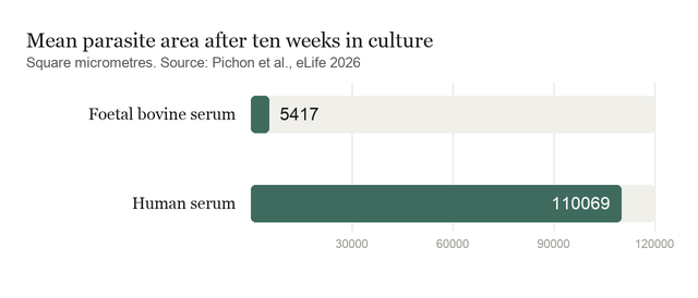
By week two, 36% of worms in human serum had reached the early liver stage against 14.8% in cow serum, and from there the paths separated completely. Cow-serum parasites rarely went further, with fewer than 0.1% reaching the late liver stage, and about three quarters were dead by week ten. Human-serum parasites moved through every stage, showed clear sexual dimorphism from around day 42, and in some experiments stayed alive beyond 150 days. Measured by area, they ended up roughly 20 times larger.
The likely reason is feeding. Schistosomes in a host begin eating blood cells around day ten, and the team added human red cells at day 13. Worms in human serum swallowed and digested them within a day, visible as hemozoin, the dark crystalline residue of haemoglobin breakdown that also accumulates in malaria parasites. Only 3.6% of cow-serum worms managed it. The interpretation is that human serum permits the gut lining to differentiate properly, and a worm that cannot digest blood cannot fuel the rest of its development.
Underneath that sits a proliferation difference visible far earlier. Labelling dividing cells showed a gap by two days after transformation, a point at which only a small defined set of somatic stem cells is dividing at all. Cow-serum parasites plateaued at no more than about 20 dividing cells per worm from day eight onward. Growth in human serum tracked stem-cell proliferation from the start.
Anatomy, pairing and the wall they hit
The worms are not merely larger. Confocal imaging found males with a gynaecophoric canal, three to five testis lobes and sperm, and females with primordial ovaries, oviduct, ootype and uterus. Sexing by PCR confirmed the dimorphism was genetic, and males and females reached it on the same schedule.
Pairing was another matter. Dish-raised worms paired with each other only rarely, only in cultures past 80 days, and only temporarily. Paired with adult worms perfused from infected mice, they coupled within 24 hours, and dish-raised females so paired began maturing their ovaries while unpaired ones stayed immature. So the machinery works; something about two dish-raised worms together does not.
What the study can't say yet
The end point is missing. Across more than 30 experiments in which worms became dimorphic and sometimes paired, not one egg was produced. Since eggs lodged in tissue cause the pathology of schistosomiasis, the part of the life cycle most worth studying is still out of reach, and the authors say plainly that further refinement is needed.
The variability is wide. Standard deviations frequently approach or exceed the means, with sexual dimorphism reached by 13.4% of worms plus or minus 18.6, reflecting the developmental asynchrony seen in real infections but also meaning any single culture may behave unlike the average. The stem-cell explanation is an inference from proliferation counts and prior single-cell work, not from transcriptomics done here.
Nor is human serum a clean win. The field moved to foetal bovine serum in the 1980s for reasons that have not gone away: batch variability, ethical sourcing and bloodborne viruses. Which molecules matter is unknown, and the authors flag both problems at once, noting that HS contains essential host-specific molecules that are absent or insufficient in FBS, and future efforts should aim to identify these factors. The sharpest implication points backward at the literature: These findings may raise some concerns about studies that rely on in vitro culture protocols using FBS, including those focused on parasite developmental biology, interaction with the host and drug screening. A compound screened against a stalled larva may say little about an adult worm.
Quick questions
Why does growing them in a dish matter? Sexual maturation currently requires infecting an animal, which limits what can be observed and manipulated. A dish culture makes the step accessible to drugs, gene knockdowns and imaging, and reduces animal use.
Is hemozoin the same thing as in malaria? Chemically yes. Both parasites eat haemoglobin and must neutralise the toxic haem released, crystallising it into an inert pigment. Here its appearance is the visible sign that a worm's gut has become functional.
What's the one-line takeaway? Swapping cow serum for human serum lets schistosomes develop into males and females entirely in culture, about 20 times larger and able to digest blood, though they still do not lay eggs.
Pichon R, Lotkowska ME, Bulathsinghalage JLD, et al. "In vitro sexual dimorphism establishment in schistosomes." eLife, 2026;15. doi.org/10.7554/eLife.111066
PubMed PMID: 42720975.
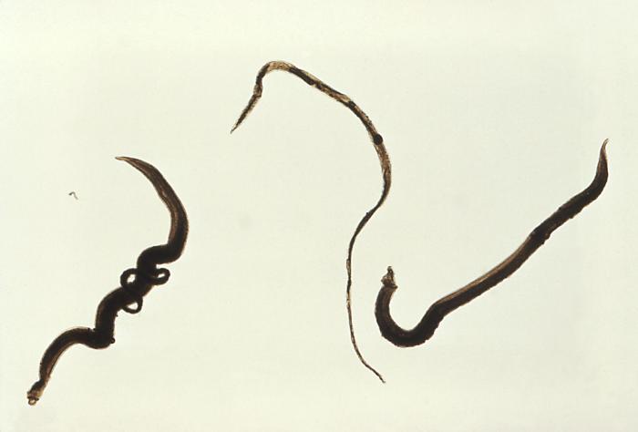
Image: Schistosoma mansoni trematodes. CDC, public domain, via Wikimedia Commons.
Comments