Autism genetics has a scale problem. Sequencing has produced a long list of high-risk genes, but they encode proteins doing visibly unrelated jobs: chromatin remodellers, synaptic scaffolds, ion channels. If each one causes the condition by its own route, there is no shared target to aim a treatment at. The open question is whether the routes converge somewhere downstream, and answering it requires a human model of cortical development rather than a mouse.
A team at Yale took one gene, DEAF1, and followed it from DNA binding to functioning tissue. DEAF1 is unusual among autism risk genes in that it encodes a sequence-specific transcription factor, so its direct targets can be listed rather than inferred. Introducing a frameshift mutation into human embryonic stem cells and growing them into cortical organoids produced a consistent picture: progenitor cells accumulated instead of becoming neurons, the cortical layers formed in the wrong proportions, and the neurons that did form fired less and connected poorly. Reanalysing published data on 36 other high-risk autism genes, the authors found that 27 of them produced the same progenitor excess, through the same upregulated pathways.
Why it matters: A convergent mechanism is the difference between needing a therapy per gene and needing one per pathway. The authors show a drug already in clinical development pushes the organoids partway back.
The study, by Kim et al., appears in Science Advances. Two details of its construction deserve attention. First, mapping where DEAF1 binds in stem cells returned 505 genes, roughly 70% of them in already-open chromatin, dominated by terms like synapse organisation and forebrain development. Second, making the mutant line was extraordinarily hard: of 384 screened clones, exactly one carried a heterozygous loss-of-function edit in the SAND domain, which the authors read as a sign of how essential the gene is in early development.
Progenitors that will not move on
The mutation closed chromatin at 1,667 genes and opened it at 4,708. The closed set is the informative one, enriched for axonogenesis, neuron migration and forebrain development, and only partly explained by direct DEAF1 binding, meaning most of the effect is indirect.
In organoids the consequences appeared once neurogenesis began. Mutant organoids were smaller by day 27 and more so by day 70, yet their ventricular zones, the proliferative layer, were larger and more densely stained for the proliferation marker KI67. Dividing radial glia shifted toward the spindle orientation associated with symmetric self-renewal, making two progenitors rather than a progenitor and a neuron. Intermediate progenitors fell. Deep-layer neurons were overrepresented and superficial-layer neurons underrepresented. The tissue was building the factory and not the product.
Function followed structure. Calcium imaging showed lower activity amplitude and frequency. Electrode arrays showed longer, more scattered bursts and poorer synchronisation between channels. Individual neurons had fewer dendritic branches and shorter dendrites, and when two organoids were fused, or linked through a 150-micrometre microchannel on a custom chip, mutant organoids projected far fewer axons into their partner.
Where the genes meet
Transcriptionally the organoids matched human fetal cortex between 8 and 24 post-conception weeks, which is the window this model can speak to. Mapped against risk genes for six neuropsychiatric and neurodegenerative conditions, the mutant profile correlated most strongly with autism, at 0.82.
The convergence claim rests on reanalysis rather than new experiments. Taking single-cell data from a published platform that perturbed 36 high-risk autism genes in organoids, the authors found that 27 of the 36 significantly increased progenitor populations relative to control. The pathways upregulated in their own mutant, WNT signalling, TGF-beta superfamily signalling and cell-cycle regulation, were the ones shared. As they write, the work revealed a striking convergence in the dysregulation of WNT signaling, TGFβ superfamily signaling, and cell cycle regulation, leading to aberrant NPC expansion.
That suggested a target. Screening clinical-stage kinase inhibitors, the team found that zamaporvint, which blocks Porcupine and so prevents WNT ligands from being secreted at all, restored much of the balance when applied at 200 nanomolar during the narrow window of radial glial expansion. Proliferation markers dropped to control levels, division angles normalised, and neuron numbers partially recovered. The choice of target is deliberate: blocking ligand secretion is more selective than suppressing the whole downstream cascade, which development depends on.
What the study can't say yet
The rescue is explicitly partial. Genes for neuronal activity and synaptic function did not recover. The authors state plainly: However, neuronal function-associated genes were not rescued, indicating that recovery remained incomplete. Normalising how many neurons are made is not the same as making them work.
The genotype does not match the patients it most resembles. The edit is heterozygous, but the organoid phenotypes, microcephaly in particular, look like those of people carrying biallelic variants. DEAF1 protein fell below half of control, leaving open a dominant-negative effect. Everything rests on a single clone from a single stem cell line; off-target sites were sequenced and came back clean, but the authors still call for patient-derived cells carrying natural mutations.
The convergence finding inherits the limits of the dataset it reuses, where cell numbers per perturbation varied, and "increased progenitor population" is a coarse phenotype that several mechanisms could produce. An organoid corresponds to early fetal cortex with no sensory input, no vasculature, no immune cells and no behaviour, so nothing here connects a cell-division angle to an autistic trait. Zamaporvint worked in a six-day window in a dish; WNT inhibition during human brain development is not a therapeutic proposition.
Quick questions
What is a cortical organoid? A three-dimensional tissue grown from stem cells that reproduces the layered organisation and cell types of early fetal cortex. It models development, not a working brain.
Why does a progenitor excess matter? Cortex is built on a schedule. Progenitors that keep copying themselves instead of differentiating produce fewer neurons of the right type at the right time, which shows up later as disrupted layers and sparse connections.
What's the one-line takeaway? A mutation in one autism gene stalls human cortical organoids at the progenitor stage, 27 of 36 other high-risk genes do the same thing through shared WNT and cell-cycle dysregulation, and blocking WNT secretion restores the cell balance but not neuronal function.
Kim J, Choe MS, Yang WS, et al. "Elucidating the role of DEAF1 in neurodevelopment and shared molecular pathways in high-risk autism genes using cortical organoids." Science Advances, 2026;12(37):eady5166. doi.org/10.1126/sciadv.ady5166
PubMed PMID: 42726852.
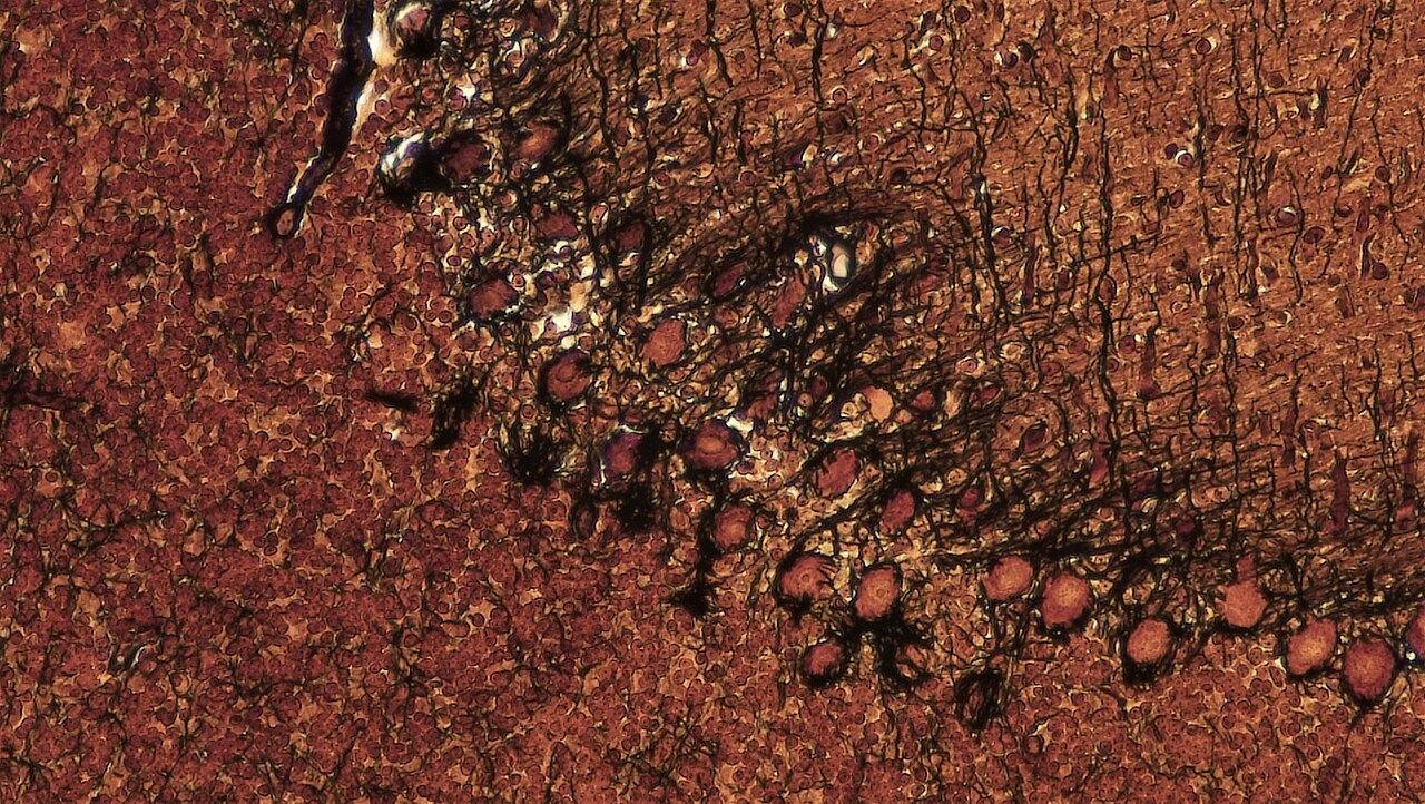
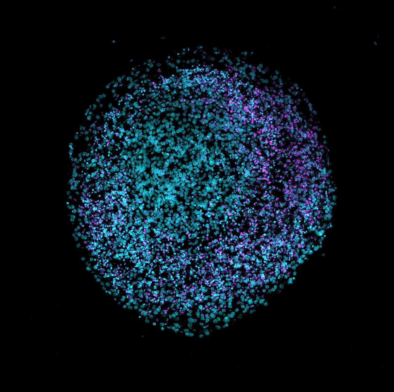
Image: cryosection of a human brain organoid. Nreis1, CC BY 4.0, via Wikimedia Commons.
Comments