The claim that an ageing cerebellum causes an older person's movements to become slower and less accurate has always rested on a weak link. The cerebellum does shrink; older adults do move less well. But the tasks used to connect the two, balance, gait, inter-joint coordination, general motor learning, all draw on muscle strength, proprioception, vision and attention as much as on the cerebellum. A decline in any of them looks the same from the outside.
A group at KU Leuven built a test battery designed to break that ambiguity. Across 160 adults, 50 young, 80 aged between 55 and 70, and 30 over the age of 80, they ran seven motor tasks and one cognitive task, and from each one extracted two separate outcomes: a measure chosen because it is specifically impaired in patients with cerebellar degeneration, and a general sensorimotor measure from the same task that is not. The two classes of measure came apart almost completely. General sensorimotor performance declined with age, as expected. The cerebellar-specific measures, in most tasks, did not, including in the participants over 80. Their cerebellums had visibly shrunk all the same.
Why it matters: If the functions the cerebellum governs hold up while the structure degenerates, then treating age-related motor problems as cerebellar atrophy is aiming at the wrong target, and the resilience itself becomes the thing worth studying.
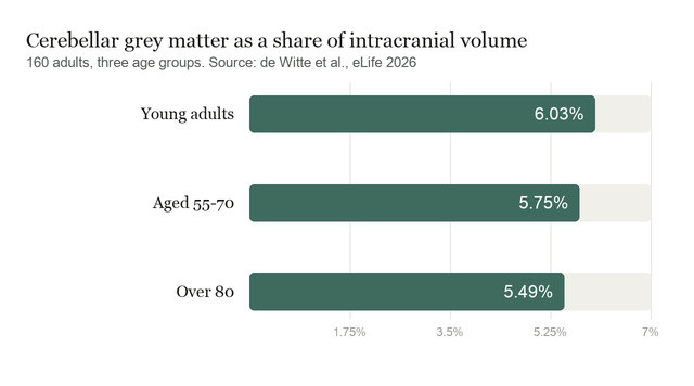
The study, by de Witte et al., appears in eLife. Structural loss in the sample was real and graded. Cerebellar grey matter, expressed as a share of total intracranial volume, fell from 6.03% in young adults to 5.75% in the 55 to 70 group and 5.49% in those over 80, a difference from young to oldest with an effect size of 1.47. White matter followed the same pattern. Against the Cam-CAN reference dataset of 653 scans spanning ages 19 to 89, these volumes sat within the expected range for each age.
Two measures from one task
The design is what makes the result readable. In the force-matching task, participants reproduced a force applied to one finger, once by moving a slider and once by pressing directly. Pressing directly engages the cerebellum's prediction of the sensory consequences of one's own action, which cancels part of the sensation, so people overshoot. That overshoot is the cerebellar measure, and it grew with age rather than shrinking: essentially zero in young adults, 0.31 newtons in the older group, larger still in those over 80. Sensitivity to force differences in the slider condition, the general measure from the same task, declined.
Reaching told the same story twice over. Baseline accuracy fell steadily with age, from a mean error of 0.55 degrees in young adults to 1.96 degrees in the oldest group. Implicit adaptation to a perturbation, the cerebellar measure, was numerically larger in both older groups than in the young, and Bayes factors of 0.079 and 0.105 supported the absence of a difference rather than merely failing to find one. Speech adaptation behaved the same way, while baseline vocal variability in the over-80 group was markedly higher. Rhythmic tapping, grip force and inter-joint coordination fit the pattern.
Two cerebellar measures did decline. Coordination between saccades and smooth pursuit was less precise in older adults, with a regression slope of 0.77 against the young adults' 1.02, where 1 is optimal. And mental rotation, the one cognitive task, slowed with age; accuracy also dropped in the over-80 group, to 81% from roughly 93% in both younger groups. Choice reaction time, a general measure, showed the largest age effect in the entire study, nearly doubling from 0.78 seconds to 1.4 seconds.
Reserve, or simply less damage
The authors argue for a cerebellar reserve: local circuit reorganisation and synaptic plasticity that keep function intact as tissue is lost. They set against it the alternative that cortical regions compensate, and they think that unlikely, because the cortical sensorimotor measures were the ones that declined, and because cerebellar circuitry is not readily taken over by other regions. As they put it, That is, while a cerebellar reserve might exist, it is sufficient to preserve cerebellar function but not all aspects of sensorimotor function.
A quieter number complicates the framing. Measured against total brain grey matter rather than skull volume, the cerebellum's share rose with age, from 12.74% in young adults to 13.67% in the oldest group. The cerebellum is shrinking, but more slowly than the rest of the brain. Part of what looks like resilience may be a structure that is simply being spared relative to the frontal and subcortical regions around it. The authors say as much, and suggest the cerebellum may be compensating for deficits elsewhere rather than the reverse.
What the study can't say yet
This is a cross-sectional comparison of three age groups, not a longitudinal one, so nothing here tracks an individual's decline. Participants were screened volunteers, right-handed, non-smoking and in good health, and the older groups had to score at least 23 on a 30-point cognitive screen. The authors chose that reduced threshold deliberately, to avoid a sample of only the highest performers, and note that six people were excluded on it.
The over-80 group is the most interesting and the least complete. They did not perform the eye-movement task at all, because of tracking problems with deep-set eyes and drooping lids, and only 20 of 30 were scanned. The label "cerebellar-specific" is itself an inference drawn from what cerebellar patients fail, not a measurement of cerebellar activity, and the authors are direct that for two of their outcomes the patient evidence is thin. No functional imaging was collected, so the reserve hypothesis is not tested here, only argued.
Finally, an absence of decline in a cognitively healthy, mobile, volunteering sample says little about people with neurological disease, and the two measures that did decline leave open whether cerebellar cognition ages differently from cerebellar motor control.
Quick questions
Does this mean the cerebellum stops mattering in ageing? The opposite. It means the motor problems older adults report are more likely to come from muscle, sensory and cortical decline, with the cerebellum among the systems holding steady.
Why test mental rotation in a motor study? Patients with cerebellar degeneration rotate mental images more slowly, so the task probes whether the cerebellum's non-motor contributions age the same way its motor ones do. Here they did not.
What's the one-line takeaway? Across eight tasks and 160 adults, measures isolating cerebellar function held up into the ninth decade while general sensorimotor performance fell, even though cerebellar grey matter shrank from 6.03% to 5.49% of intracranial volume.
de Witte A, Matthijs A, Parrell B, et al. "Preserved cerebellar functions despite structural degeneration in older adults." eLife, 2026;15. doi.org/10.7554/eLife.109440
PubMed PMID: 42703946.
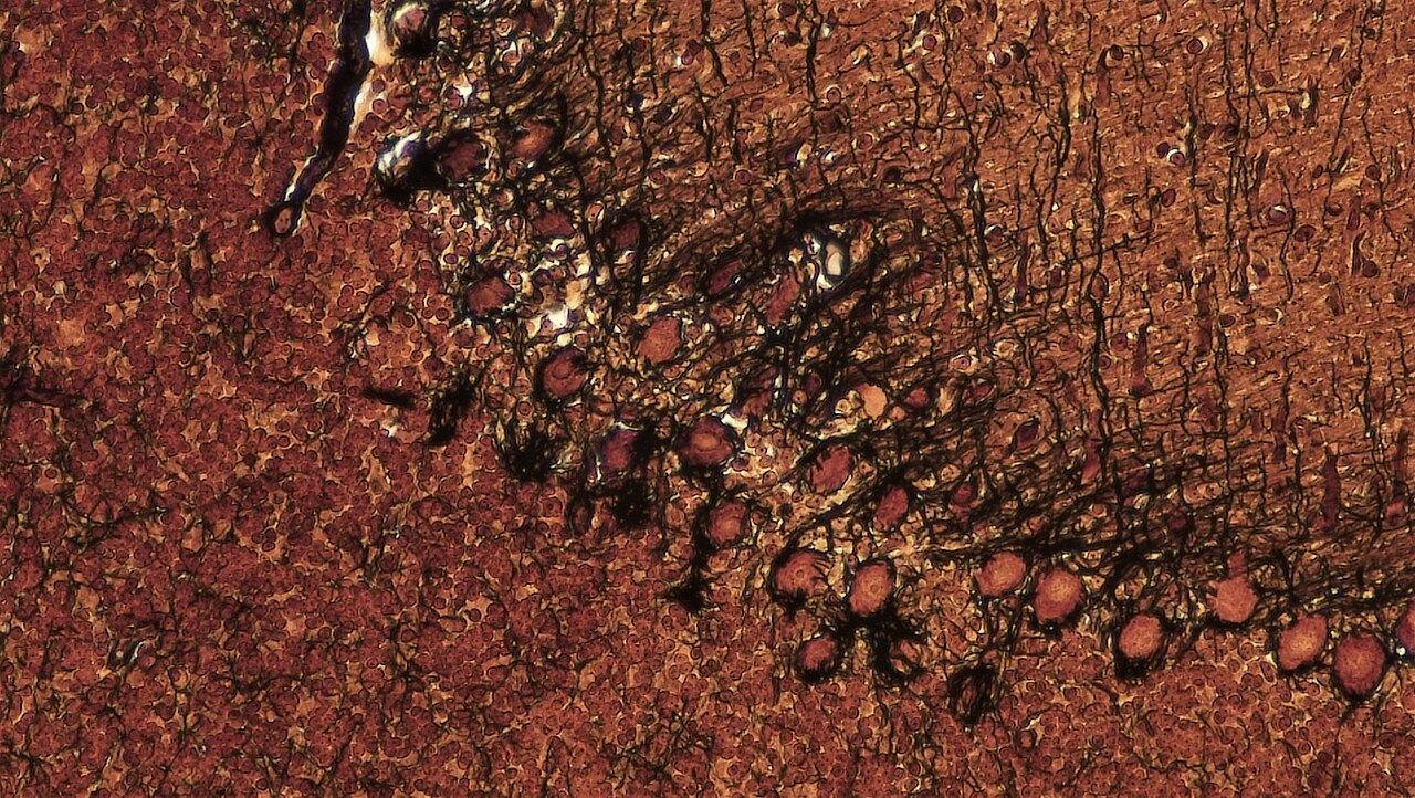
Image: cerebellum cross section, Purkinje cells, silver impregnation. Berkshire Community College Bioscience Image Library, CC0, via Wikimedia Commons.
Comments