

A century and a half of studying language in the brain has produced a lot of disagreement, and two of the reasons are methodological rather than substantive. The first is that researchers do not agree on what language is, so different labs define the target differently and localize different things. The second is that language areas sit in slightly different places in different people, so averaging across a group smears the boundaries until the network dissolves into the tissue around it.
Work at Stanford and MIT has now tested a way around both. Rather than asking where the brain lights up during a language task, individualized functional connectomics asks which parts of a single brain fluctuate together over time, and lets the networks fall out of the correlations. Applied to 1,957 scanning sessions from 1,199 people performing an assortment of unrelated tasks, the procedure kept producing a left-lateralized frontotemporal network that behaves like the language network, without ever being told to look for one.
Why it matters: A structure you can recover from arbitrary imaging data, with no language task and no theory of what language is, is on much firmer ground than one that only appears when you go looking for it with a particular definition in hand.
Deriving a network instead of testing for one
The procedure is deliberately uninformed. It takes the slow fluctuations in the blood-oxygen signal, correlates each small volume of grey matter against a set of reference regions, and clusters the result. No task structure enters the computation. The authors did not even regress the task out of the timecourses, which they note is the simpler and more general choice, and they report that a comparable network emerges when task regression is applied.
Because clustering depends on where it starts, they ran it a hundred times from random initializations per session and combined the results into a probabilistic parcellation. That converts a brittle single answer into a distribution, and it is the kind of methodological care that determines whether a result at this scale means anything.
Four properties that make it convincing
The resulting network is left-lateralized in most brains and sits mainly in the temporal and frontal lobes, which is where decades of task-based work had already put it. Agreement with expectation is reassuring but weak evidence on its own. Three further properties do more work.
It is selective, and it doubly dissociates from the networks immediately adjacent to it, meaning each responds to conditions the other does not. Adjacency in the cortex is exactly where spurious merging happens, so separating two neighbours in both directions is a strong specificity claim. It is robust to parcellation granularity: asking the algorithm for more or fewer networks does not fragment it, which implies the network is tightly integrated rather than an artefact of where you draw the lines. And it is stable across sessions within a person while differing between people, which is the signature of individual anatomy rather than noise.
The practical consequence
If the language network can be extracted from any functional run, then an enormous quantity of existing imaging becomes usable for language research retrospectively. The authors put their finger on a specific and familiar frustration, noting the value of the approach in the unfortunately common case where the task parameters associated with a given run (e.g., in an open dataset) are missing, incomplete, or poorly documented. As Shain and Fedorenko observe in Nature Communications, that covers a great deal of what has been publicly shared.
The clinical version matters more. Presurgical mapping before tumour or epilepsy surgery exists to avoid cutting language cortex, and it currently requires a cooperative patient performing language tasks in a scanner. A method that works from arbitrary scans would extend that to patients who cannot comply.
What the study can't say yet
Functional connectivity is correlation in a slow haemodynamic signal, not communication. Regions fluctuating together may be talking, may be listening to a common source, or may share a vascular quirk, and this design does not distinguish those. Calling the result a network is a statement about statistical structure.
The selectivity claim also inherits a definition of language from whatever localizer contrasts were used to evaluate it. The method sidesteps theoretical commitments while deriving the network and then reintroduces them at the point of asking whether the network is language-selective, which is a narrower escape from the definitional problem than it first appears. And demonstrating that a network recoverable without a task resembles the one found with a task is not the same as demonstrating it is identical, which for surgical planning is the distinction that would matter.
Quick questions
Does this mean fMRI language tasks are unnecessary? Not yet. The task-free network matches the task-based one in topography and behaviour, and the agreement has not been validated to the standard a surgical decision requires.
Why does individual variability cause so much trouble? Because averaging brains whose language areas sit in slightly different places blurs the boundaries until the network stops looking like a discrete structure.
What's the one-line takeaway? Across 1,199 brains doing unrelated tasks, clustering resting fluctuations alone kept yielding a left-lateralized frontotemporal network that is language-selective, stable within people, and separable from its neighbours.
Shain and Fedorenko. "A language network in the individualized functional connectomes of 1199 human brains doing arbitrary tasks." Nature Communications, 2026;17(1). doi.org/10.1038/s41467-026-75745-8
PubMed PMID: 42595752.
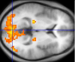
Image: Functional magnetic resonance imaging, public domain, via Wikimedia Commons.
Comments