Anti-inflammatory drugs have repeatedly failed to slow Alzheimer's disease, and part of the reason may be that "neuroinflammation" is not one thing. Microglia, the brain's resident immune cells, adopt many distinct transcriptional states in a diseased brain, and the field has no dependable way to tell which are dismantling tissue and which are holding the line. In a patient every state arrives at once, alongside amyloid plaques, hyperphosphorylated Tau and dying neurons, so injury and response cannot easily be pulled apart.
A hibernating rodent offers a way around that. Putting golden hamsters through full cycles of torpor and arousal, a Belgian group sequenced the nuclear transcriptome of around 100,000 hippocampal cells and found that microglia shifted more than almost any other cell type, entering inflammatory states that align closely with those catalogued in Alzheimer's patients. The hamsters had no detectable amyloid plaques and no neurodegeneration, and the disease states that matched were the early ones.
Why it matters: If the microglial profiles that appear first in Alzheimer's are ones a hibernating brain switches on and later switches off without lasting damage, then suppressing inflammation wholesale may remove a protective response rather than a pathological one.
The study, published in Science Advances by Largo-Barrientos et al. at the VIB-KU Leuven Center for Neuroscience in Belgium, is, its authors say, the first single-cell atlas of a hibernating brain. The link to dementia is not new: hibernating animals accumulate hyperphosphorylated Tau at sites associated with Alzheimer's disease, reversibly and without brain damage. What was missing was cell-type resolution.
What torpor does to a hippocampus
Three-month-old male golden hamsters were moved into a hibernaculum where temperature was wound down to 4°C and the lights to full darkness, with movement detectors marking the phases. Torpor bouts averaged 4 to 5 days, arousal bouts 1 to 2 days. Animals were killed in the first hours of torpor or arousal, alongside euthermic controls, across three experimental repeats; 34 cell clusters were annotated.
Neurons and glia then diverged informatively. Neurons rewrote their transcriptome heavily during torpor, with over 3,000 genes changed in common across granule cells and CA1 and CA3 pyramidal neurons, and in arousal returned to a state the authors call highly similar to control. Glia did not go back. They acquired an entirely new signature more closely related to torpor than to control, with microglia and astrocytes shifting most. Mitochondrial complex I subunits and ribosomal genes both rose in torpor, which is counterintuitive given that translation in a torpid brain is reported to fall; the authors read the ribosomal rise as preparation for arousal.
Which Alzheimer's states match, and which do not
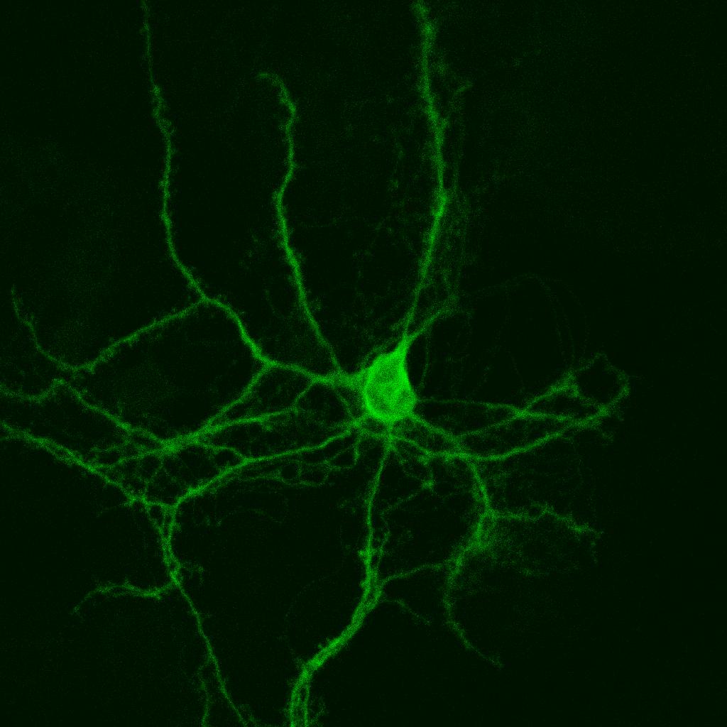
Microglia sorted almost cleanly by phase into control, torpor and arousal subclusters, plus a dividing population 67% composed of cells from aroused animals. Torpor microglia expressed ribosomal and iron-storage genes; arousal microglia expressed pro-inflammatory genes and the cytokines Ccl3 and Il1b. Morphology tracked transcription: processes in torpid animals were shorter and thinner, and significantly more of those cells carried lipid droplets, a proinflammatory feature tied to ageing and to cognitive decline in Alzheimer's disease.
The comparison to human disease was made by aligning hamster microglia against published human microglial states with SAMap and Spearman rank correlation. The torpor state matched a human state defined by ribosome biogenesis and iron-storage genes, and correlated with phagocytic and stress-response states. The arousal state matched homeostatic microglia and an inflammatory state that, in the human data, shows its greatest response in early Alzheimer's disease. The two inflammatory states that respond at late stages of the disease did not match either hamster state well. Amyloid plaques, meanwhile, were undetectable in hibernating and control hamsters, while AT8-positive phosphorylated Tau did accumulate in torpor and arousal.
How much weight the comparison can carry
One methodological choice deserves credit. Rather than treating each nucleus as an independent observation, which inflates significance through pseudoreplication, the authors aggregated counts into pseudobulk samples by cell type, condition and repeat, giving 3 to 4 samples per condition and cell type. The microglial enrichment analysis used 2 to 4 pseudobulk samples per animal, with 3 to 4 animals per condition across three conditions: a small number, normal for this kind of work and still a limit on claims about variation between individuals.
The weaker link is the cross-species comparison itself, which rests on alignment scores and rank correlations between datasets, not on an experiment where a hibernation state was induced and a disease outcome measured. The aging and injury analysis shows why that matters: formal correlation and gene-set enrichment found no significant associations with aged or injured mouse microglia, yet marker modules for those conditions were strongly upregulated in the torpor and arousal states. The authors report both, and the honest reading is that "resembles" means overlapping marker expression rather than demonstrated equivalence of function.
What the study can't say yet
Nothing here shows the hibernation states are protective. That is inferred from three observations: the states resemble early rather than late disease states, they are reversible, and they occur without lasting damage. No one blocked them to see what broke. The animals were all young males, so neither sex nor age is addressed, and a hibernator may carry safeguards a human brain lacks. The readouts are transcripts, protein levels and cell morphology, not cognition. Reversibility rests on timing too: astrocytes had not regained their control distribution 24 hours into arousal, and recovered fully only after at least a week euthermic. The authors are explicit that the protective reading remains a hypothesis, writing that Further research will be needed to confirm this hypothesis and determine whether hibernation can serve as a model to dissociate protective from detrimental neuroinflammatory responses relevant to AD.
Quick questions
Do hibernating hamsters get Alzheimer's disease? No. They accumulate the phosphorylated Tau associated with it and mount microglial responses resembling it, then clear both. That dissociation is the point of using them.
Does this mean inflammation is harmless in Alzheimer's? No, and the pattern argues the opposite: the late-stage inflammatory states, which the authors suspect are the damaging and irreversible ones, are exactly what hibernation does not produce.
What is the one-line takeaway? A hibernating hamster's brain turns on something resembling early Alzheimer's inflammation and turns it off again, offering a way to study that response apart from the disease.
Largo-Barrientos et al. "Common transcriptional changes in the brain during hibernation and Alzheimer's disease." Science Advances, 2026. doi.org/10.1126/sciadv.aeg3486
PubMed PMID: 42981396.
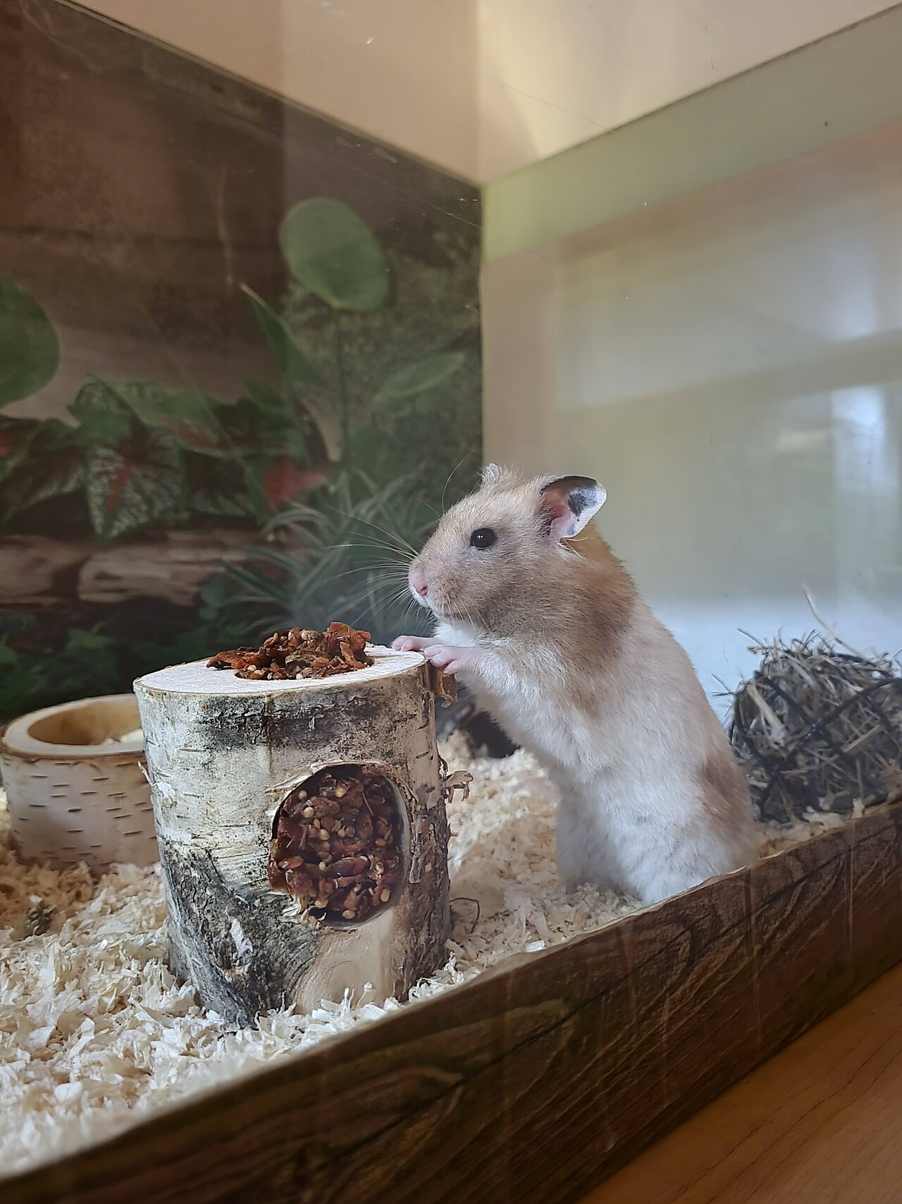
Image: a golden hamster (Mesocricetus auratus), the species used in the study. Patrik Čech, CC BY-SA 4.0, via Wikimedia Commons.
Comments