

Patients on haemodialysis need reliable access to their bloodstream, usually through a surgically created connection between an artery and a vein. These arteriovenous fistulas fail often, because the vein wall thickens and narrows. Understanding what drives that thickening is a real clinical problem, and a 2024 paper proposed an answer: a long non-coding RNA that pushes smooth muscle cells into a proliferative state.
A group from Edinburgh, Maastricht and the Weizmann Institute has now published a formal objection, and it is not about the biology. It is about whether the RNA in question exists as a separate thing at all.
Why it matters: The proposed RNA sits entirely inside an intron of a protein-coding gene, on the same strand, and is unspliced. In that configuration, a signal from the region is genuinely ambiguous. It could come from an independent transcript, or it could come from the unfinished pre-mRNA of the host gene as it is being made.
Why the configuration is the problem
Most genes are transcribed as a longer precursor from which introns are cut out. So the intron sequence does exist as RNA, briefly, inside every cell expressing that gene. If you design an experiment that detects that sequence, you will get a signal whether or not a separate RNA is also produced there.
Spliced RNAs avoid this trap: reads spanning the joins between their own segments are unique to them. An unspliced RNA lying inside an intron has no such signature, and would share complete sequence identity with the host precursor. The critics argue that this puts the burden of proof squarely on anyone claiming the transcript is real. As Bennett and colleagues write in Nature Communications, Intronic lncRNAs require unambiguous evidence to show they are a distinct transcriptional unit with independent transcriptional initiation and termination. Some annotation projects handle this by excluding unspliced transcripts entirely rather than risk cataloguing artefacts.
What they looked for and did not find
The group examined public total RNA sequencing from samples matching those in the disputed work, including mouse heart, and human coronary artery and aortic smooth muscle cells from ENCODE. If a distinct transcript were produced at that spot, reads should pile up there relative to the surrounding intron. They report no such enrichment: coverage at the site was no higher than the low background found across the whole intron.
They also checked for a promoter using CAGE, a method that maps where transcription starts. An independent gene needs its own start site. They found no robust hits anywhere in the intron.
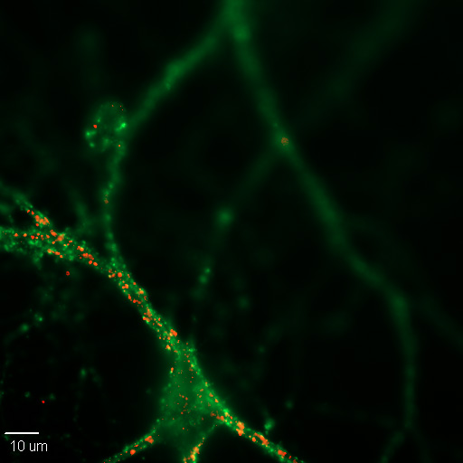
The sharpest point is an internal inconsistency. The 2024 paper's imaging shows an intense signal in the cytoplasm, which would indicate an abundant, exported transcript. Something that abundant should be conspicuous in sequencing data from the same tissues. It is not there. The authors add that they could not locate the sequence of the imaging probe in that paper or its predecessors, which makes the discrepancy impossible to chase down.
How this propagated
The critics trace the original assignment to a 2014 microarray study, and note a diagnostic feature of it: it reported that 55 percent of the long non-coding RNAs it identified were intronic, against 2.5 percent in a stringent contemporary annotation. A catalogue where intronic entries dominate to that degree is more likely picking up precursor signal than discovering an unusual class of genes.
That matters because five studies now rest on the assignment, none of which, in the critics' reading, went back and established it. If the target is really the host precursor, then the interventions used across those papers, which include overexpression, RNA interference and conditional knockouts in mice, may have been altering the protein-coding gene rather than a separate RNA. The critics note that the host gene is not mentioned in the 2024 study at all, and that where its levels were reported in earlier work, the analyses were underpowered.
What the study can't say yet
Absence of evidence in existing datasets is not proof of absence. The transcript could be produced in a cell state or condition not represented in the sequencing they examined, or at a level that public data cannot resolve. The critics say only that they cannot find support for it, which is a weaker and more accurate claim than saying it is not there.
Nothing here shows the biological results are wrong. The vein-thickening phenotypes reported in those papers may well be real and reproducible. What would change is the explanation: the same experiments would then be evidence about a protein-coding gene, and would need reinterpreting rather than discarding.
This is also one side of an open exchange. Formal objections of this kind are published alongside a reply, and resolving it requires either the original authors producing the missing annotation evidence or someone running the specific tests, such as mapping the transcript's ends, that would settle it directly.
Quick questions
What is a long non-coding RNA? An RNA transcribed from the genome that is not translated into protein. Many have real regulatory roles, which is why claims about new ones are taken seriously and why misassignments are costly.
Why can't you just measure it? Because it would be identical in sequence to part of the host gene's precursor. Distinguishing them needs evidence about where transcription starts and stops, not simply whether the sequence is present.
What's the one-line takeaway? An RNA underlying five studies on blood vessel remodelling shows no independent read coverage and no promoter signal in public data, raising the possibility that experiments targeting it were targeting the surrounding gene instead.
Bennett et al. "In search of LncDACH1 - a perceived intronic long non-coding RNA lacking fundamental genomic annotation evidence." Nature Communications, 2026;17(1). doi.org/10.1038/s41467-026-76991-6
Responding to: Li et al. Nature Communications, 2024. doi.org/10.1038/s41467-024-48019-4
PubMed PMID: 42629347.
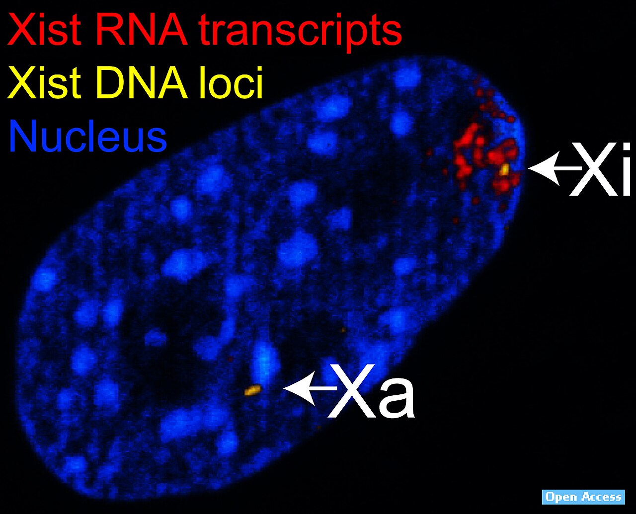
Image: RNA and DNA fluorescence in situ hybridisation, B. Reinius and C. Shi, CC0, via Wikimedia Commons.
Comments