
The textbook divides brain chemistry in two. Fast transmitters such as glutamate cross a synapse, a junction a few tens of nanometres wide, from one named partner to another. Neuromodulators such as dopamine are supposed to work the other way: released from swellings along an axon that have no synaptic structure, drifting through the extracellular space, reaching whatever receptors they happen to meet. This is called volume transmission, and it has an awkward problem. Dopamine neuron firing shapes behaviour on timescales of milliseconds to seconds, which is difficult to reconcile with a signal that gets where it is going by diffusion.
A group at Janelia Research Campus has now watched dopamine leave individual axonal swellings and then gone back to the same spots to ask what was there. Most swellings released nothing. The ones that did were pressed directly against the cell bodies and dendrites of neurons that carry dopamine receptors, carried the protein machinery of a conventional presynaptic terminal, and sat beside tight clusters of receptors rather than a smooth carpet of them. Contact, not chance, predicted which sites worked.
Why it matters: Nearly every drug that touches dopamine, from antipsychotics to Parkinson's therapies, is designed around assumptions about where dopamine goes after release. If release is anchored opposite receptor clusters rather than broadcast, the relevant concentration is a local one, and the target is a structure rather than a volume.
The study, by Bulumulla et al. at the Howard Hughes Medical Institute's Janelia Research Campus, appears in Science Advances. It combines two nanomaterial dopamine sensors, one a film that cultured neurons grow on and one usable in brain slices, with gene expression mapping, electron microscopy, and two new lines of mice carrying a small tag knocked into the D1 or D2 receptor gene.
Most release sites do not release
Growing dopamine neurons alongside cortical or striatal neurons on the sensor film let the team see release from single swellings. Within a field of view, 34% of swellings were release-competent, with a standard deviation of 27 percentage points, so the fraction varies enormously between preparations. Active sites gave a fluorescence change of about 14.7% against 3.65% for silent ones, and the weak signals at silent sites peaked after 4.55 seconds rather than 0.81, which the authors read as dopamine drifting in from a neighbour rather than being released on the spot.
Active sites were not scattered. They clustered, peaking at interbouton distances around 10.5 micrometres, whereas silent swellings showed a featureless distribution. They also tracked a marker of dendrites and cell bodies: release amplitude fell off with distance from neuronal processes, and was highest where axon touched target directly. Proximity to glia did nothing. The specificity goes further than contact. Where dopamine axons wrapped neurons lacking receptor transcripts, they stayed silent, so touching a neuron is not enough; the neuron has to carry receptors.
Receptors sit in clusters, next to the sites that work
Dopamine receptors are notoriously hard to stain, which is why their subcellular arrangement has stayed vague. Knocking a short tag into one copy of each receptor gene solved it. D1 and D2 both appeared as discrete puncta, not diffuse membrane, averaging 0.72 and 1.05 micrometres across respectively, with D2 largely confined to dendritic shafts while D1 also reached into spines.
In intact tissue the pattern held anatomically. Mapping receptor transcripts across more than 2500 amygdala neurons and measuring how much dopamine axon wrapped each cell body, receptor-expressing classes drew between roughly 490 and 964 cubic micrometres of axon, against 78 for receptor-negative neurons. The same bias appeared in dorsal striatum. Receptor clusters apposed to a dopamine swelling were also larger than unapposed ones, which is what happens at ordinary synapses.
Release machinery followed suit. Bassoon, a scaffold that marks the active zone, was roughly three times more abundant at active than silent swellings, and knocking down Munc13-1, RIM, syntaxin 1 or synaptotagmin 1 nearly abolished release; bassoon knockdown reduced it without eliminating it.
What the study can't say yet
The word doing the work is "synapse-like", and the electron microscopy explains why. Classical synaptic structure, with parallel membranes and a clear cleft, appeared in 60% of non-dopamine terminals but only 40% of dopamine ones. Most dopamine contacts were narrow, undulating interfaces without a defined cleft. These are not ordinary synapses, and the paper does not claim they are.
Almost all the functional work is in dissociated neurons on a flat sensor film, and the authors themselves note that cultured neurons are so synaptogenic they will build synapses onto protein-coated beads. That cuts against the result as much as for it: a preparation eager to form contacts may overstate how orderly the arrangement is in a brain. The tissue experiments are anatomical rather than functional, so nobody has yet recorded release from a single identified swelling in intact tissue and then asked which receptors were opposite it.
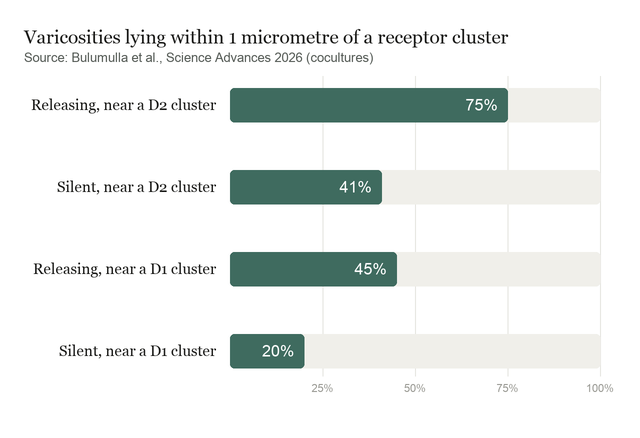
The association statistics are strong in direction and loose in magnitude. Active swellings were enriched near receptor puncta with odds ratios of 7.85 and 15.7 for D1 and D2, but the reported uncertainties of 4.8 and 10.4 are large fractions of the estimates. And the causal arrow is unresolved: knocking down release proteins shows they are needed for release, not that apposition is what recruits them. On that the authors are explicit, writing that Future work combining unbiased proteomics with targeted perturbations will be needed to establish a causal link between synapse-like appositions with presynaptic release maturation.
The three-dimensional reconstruction that shows vesicle clusters lining up with release hotspots is a single volume, and the accompanying observation about mitochondria is flagged as unquantifiable for that reason.
Quick questions
Does this mean diffusion plays no part? No, and the authors say so. Their proposal is hybrid: release anchored at receptor-proximal sites, with spillover still reaching nearby receptors, particularly when activity is high.
Why would it matter whether dopamine is targeted or diffuse? Because it changes what counts as a dose. A receptor sitting opposite a release site experiences a brief, high concentration; one a few micrometres away experiences something much weaker and later.
What is the one-line takeaway? Dopamine release is concentrated at a minority of axonal swellings that press against receptor-bearing neurons and carry conventional presynaptic machinery, shown convincingly in cultured neurons and so far only anatomically in brain tissue.
Bulumulla et al. "Synapse-like specializations at dopamine release sites orchestrate efficient and precise neuromodulatory signaling." Science Advances, 2026. doi.org/10.1126/sciadv.aei0203
PubMed PMID: 42826180.
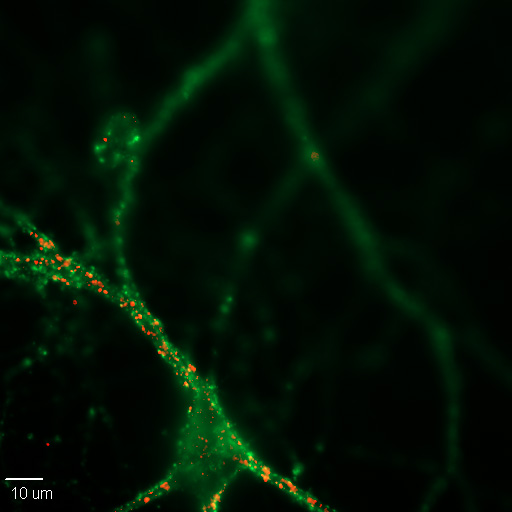
Image: cultured mouse cortical neuron with synapses labelled for synaptophysin and PSD-95. Dchordpdx, CC BY 4.0, via Wikimedia Commons.
Comments