
Computational protein design has moved faster than the ability to check what the designs do. Generating a hundred plausible variants of a protein is now routine; determining which of them behaves correctly is not, and the gap widens whenever the property of interest is a behaviour rather than a measurement. Binding affinity can be read off a plate. Whether a protein organizes itself into filaments, and how quickly, and whether those filaments bundle, cannot.
A group at the Max Planck Institute of Biochemistry has built a screen for that second category. PUREdrop encapsulates each designed protein, along with the machinery to manufacture it, inside thousands of droplets a few picolitres in volume, then distributes them across a 96-well plate and films them. Each droplet is a crude artificial cell, and what the protein does inside one is watched over 24 hours rather than measured once at the end.
Why it matters: The Design-Build-Test-Learn loop that drives protein engineering is only as fast as its slowest step, and for self-organizing behaviours the slow step has been Test. A screen that captures how a design unfolds in time closes the loop for a class of proteins it was previously open for.
Why an ordinary screen cannot see this
The authors are explicit about the deficiency they are addressing, writing that A great bottleneck of this new research direction is the availability of standardized and comprehensive screening routines that provide direct access to spatiotemporal readouts. Two words there carry the weight. Spatio, because a protein that assembles into a structure needs to be somewhere in particular, and a well-stirred tube destroys exactly the spatial information you want. Temporal, because assembly has kinetics, and an endpoint measurement discards them.
Confinement matters for a subtler reason. A filament forming in bulk solution has effectively unlimited room; inside a cell it meets a boundary at a scale comparable to its own length, and the boundary changes what forms. A picolitre droplet imposes something like that constraint, which is why the readout resembles the environment these proteins are eventually meant to work in.
What they screened, and what came back
The test case is FtsZ, the protein bacteria use to divide. It polymerizes into filaments that gather into a ring at the midpoint of the cell and constrict, and it is the obvious starting point for anyone trying to build a synthetic cell that can divide. As Al Nahas and colleagues report in Nature Communications, they ran 24 computationally redesigned variants through the platform.
Three behaved differently enough to count as hits, and two of them differed in opposite directions, which is the informative result. One variant began bundling earlier and bundled more heavily than the wild-type protein. Another began later and bundled less. A screen that only reported which variants worked would have discarded the second as a failure; a screen that reports kinetics shows it as a variant with a different, potentially useful, tempo.
They then applied the same setup to a different question, testing not variants of FtsZ but other proteins that modulate it. One combination anchored the filaments to the droplet interface and produced a ring-like arrangement, which is the geometry the real protein adopts in a dividing bacterium.
Why the timing data is the point
Bundling dynamics might read as a technical detail, and the paper makes the case that they are not. A synthetic cell has a fixed budget of transcription and translation capacity shared among everything it expresses. A variant that bundles early and heavily consumes resources on a different schedule from one that bundles late, and when several engineered protein systems have to coexist in one compartment those schedules collide. Choosing a variant on tempo rather than on endpoint performance is what makes the parts combinable.
What the study can't say yet
A droplet is not a cell. There is no membrane in the biological sense, no metabolism, no capacity to regenerate the transcription-translation machinery, which is why the experiment ends at 24 hours. What is being screened is protein behaviour under confinement, and the step from that to behaviour inside anything self-sustaining is untested here.
Twenty-four variants is also a modest library for a platform whose case rests on throughput. The architecture plausibly scales, and scaling was not demonstrated. Nor does the work explain why the successful redesigns behave as they do: the variants came from a computational method and the screen sorts them, but no structural account connects a sequence change to the altered bundling. That is the Learn half of the loop, and it remains open. The ring-like phenotype is likewise a geometry, not a division, and the distance between a filament ring and a cell that splits in two is the whole remaining problem.
Quick questions
Are these synthetic cells alive? No. They are droplets containing DNA and the molecular machinery to express it, capable of running a protein-production programme for about a day and nothing beyond that.
Why screen inside droplets rather than in a tube? Because the behaviours of interest are spatial and time-dependent. A tube gives one bulk number at one moment and discards both.
What's the one-line takeaway? An automated platform expressed 24 redesigned versions of the bacterial division protein FtsZ inside thousands of picolitre droplets each and filmed them, finding three variants whose filament bundling ran on visibly different schedules from the original.
Al Nahas et al. "Automated synthetic cell-based screening for designed proteins with emergent functions." Nature Communications, 2026;17(1). doi.org/10.1038/s41467-026-76787-8
PubMed PMID: 42603823.
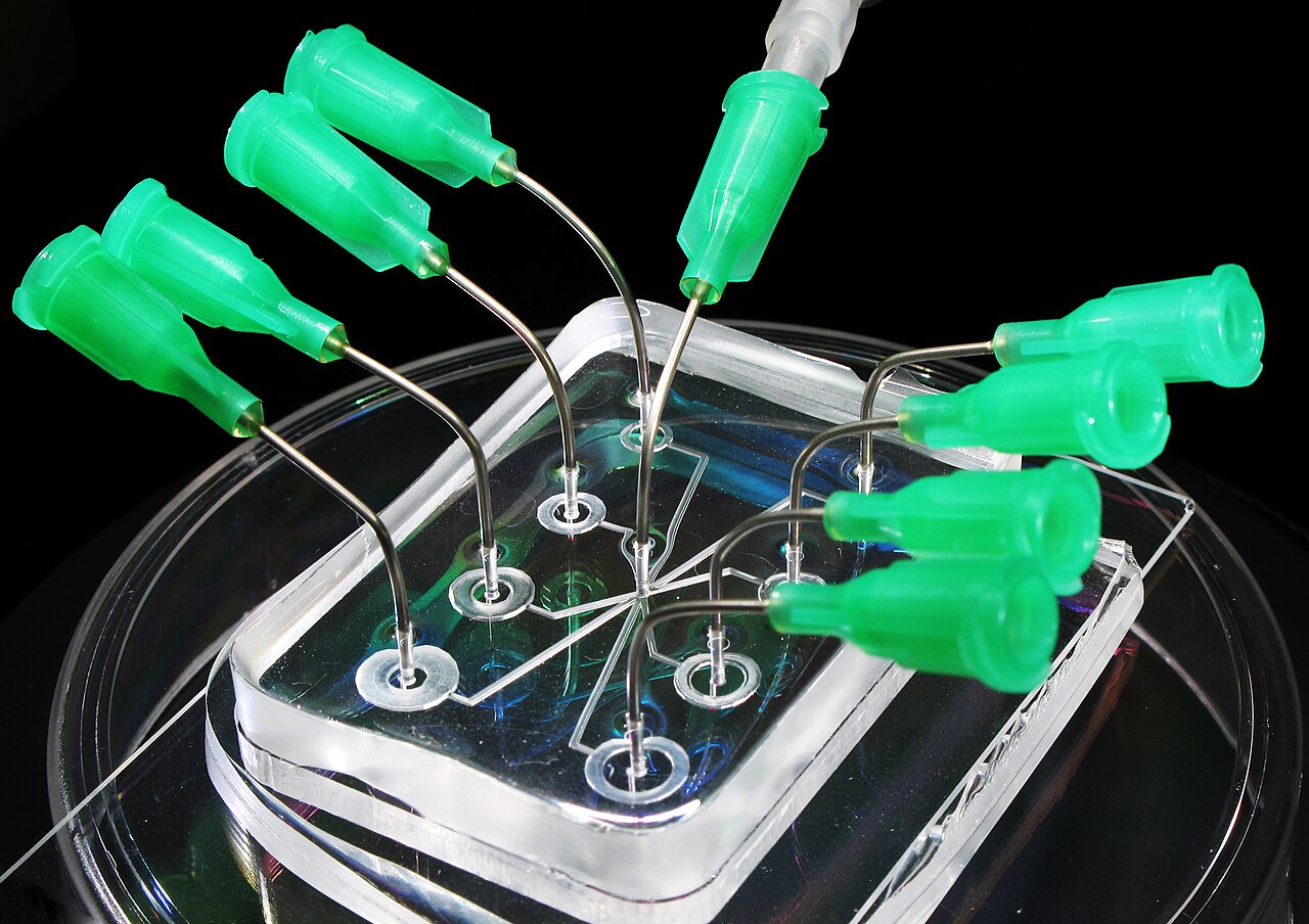
Image: Lab-on-a-chip microfluidic device, National Institute of Standards and Technology, public domain, via Wikimedia Commons.
Comments