

Retinal organoids have a persistent quality problem. Grown from stem cells, they self-organize into something with the right layers and the right cell types, and then stall. The photoreceptors never quite finish: outer segments stay stunted, gene expression hangs between precursor and mature states, and light responses are weaker than a real retina's. Every downstream use, disease modelling, drug screening, cells intended for transplant, inherits that shortfall.
A collaboration between Masaryk University and Newcastle asked whether the missing ingredient is the obvious one. A retina develops inside an animal that experiences light, and light is not merely what photoreceptors detect but a signal present throughout their development. Organoids are grown in the dark. Exposing them to light flickering at 40 hertz produced measurably more mature photoreceptors.
Why it matters: If a widely used organoid system underperforms because it is missing a physiological input rather than a molecular one, the fix is a light-emitting diode on a timer. That is unusually cheap and scalable for a quality improvement in stem cell work.
Why the frequency is the finding
The result would be far weaker without the comparison conditions, and this is where the study earns its conclusion. Constant illumination was tested. Randomly flickering light was tested. Neither did what 40 hertz did. The effect depends on temporal structure at a specific rate, not on light exposure as such, which rules out the dullest explanations, that any photic energy accelerates development or that the cultures were simply warmer.
Forty hertz sits in the gamma band, the same frequency that has drawn attention in work on rhythmic sensory stimulation elsewhere in neuroscience. Whether the two phenomena share anything mechanistically is entirely open, and the paper does not claim they do. What it establishes is narrower and firmer: among the paradigms tried, this one worked and the others did not.
What improved, at three levels
Maturation was assessed structurally, transcriptionally and functionally rather than by any single proxy. Structurally, the outer segments, the stacked membrane compartments where phototransduction happens and the feature organoids most conspicuously fail to build, elongated. Transcriptionally, expression shifted away from precursor states toward mature photoreceptor identity. Synaptic protein expression rose.
Functionally, the claim is deliberately modest. As Celiker and colleagues report in Nature Communications, the stimulation produced a modest but reproducible enhancement of light-evoked responses in downstream retinal neurons. Retinal ganglion cells, two synapses downstream of the photoreceptors, responded better to light. That the improvement is described as modest rather than dressed up is the reason to take the rest of it seriously.
What an outer segment is and why it fails
The structure that organoids struggle to build is worth understanding, because it is the one that does the actual work. A photoreceptor's outer segment is a dense stack of membrane discs, hundreds of them, packed with opsin. It is metabolically expensive, continuously renewed from the base while the tips are shed and eaten by the pigment epithelium, and it is where a photon is converted into an electrical signal. A photoreceptor without a proper outer segment is a cell of the right identity that cannot do the job.
In organoids these come out short and disorganized, which is the single clearest marker of arrested maturation and the reason light responses stay weak. So elongation under stimulation is not a cosmetic morphology score. It is movement on the specific structure whose absence explains the functional deficit, which is why the transcriptional and electrophysiological changes reported alongside it are coherent rather than a scatter of unrelated readouts.
Borrowing a principle from other organoids
The underlying logic is established elsewhere and had simply not been applied here. Cardiac organoids are mechanically stretched and electrically paced, which improves sarcomere formation and alignment. Neuronal cultures given electrical pulses develop synaptic networks faster. Pancreatic islet organoids mature better under time-restricted feeding that imposes a circadian rhythm. In each case the intervention supplies a physical input the tissue would meet in the body and does not meet in a dish.
Light is the obvious analogue for retina and was overlooked, plausibly because a retina in a dish has no eye around it and the question of what it should be exposed to has no natural answer. Animal work already pointed the way: light exposure during defined developmental windows shapes photoreceptor orientation and synaptic transmission, and dark rearing delays those processes. This study carries that into a human system.
What the study can't say yet
Mechanism is absent. Nothing here explains why 40 hertz specifically, or what transduces a flicker rate into a transcriptional programme in a cell whose phototransduction machinery is by definition still immature. Without that, the frequency is an empirical optimum from the paradigms tested rather than a biologically meaningful number, and a finer sweep might find something better or reveal a broad plateau.
Maturation is also improved, not achieved. Organoid photoreceptors remain short of the real thing on every measure reported, and the gap that matters for transplantation is whether grafted cells connect and signal in a host retina, which no amount of in-dish characterization settles. The functional gain being modest is precisely what makes the leap to therapeutic relevance premature.
Quick questions
Does this help people with retinal disease now? No. It improves the laboratory tissue used to study those diseases and to develop treatments, which is upstream of any clinical effect.
Why would a flicker rate matter when constant light doesn't? Unresolved. The comparison establishes that temporal patterning rather than illumination is doing the work, without identifying what reads the pattern.
What's the one-line takeaway? Retinal organoids grown under 40 hertz flickering light built longer outer segments, shifted toward mature photoreceptor gene expression, and produced modestly better light responses than those grown in the dark or under constant illumination.
Celiker et al. "Photostimulation improves maturation of human photoreceptors." Nature Communications, 2026;17(1). doi.org/10.1038/s41467-026-76112-3
PubMed PMID: 42586989.
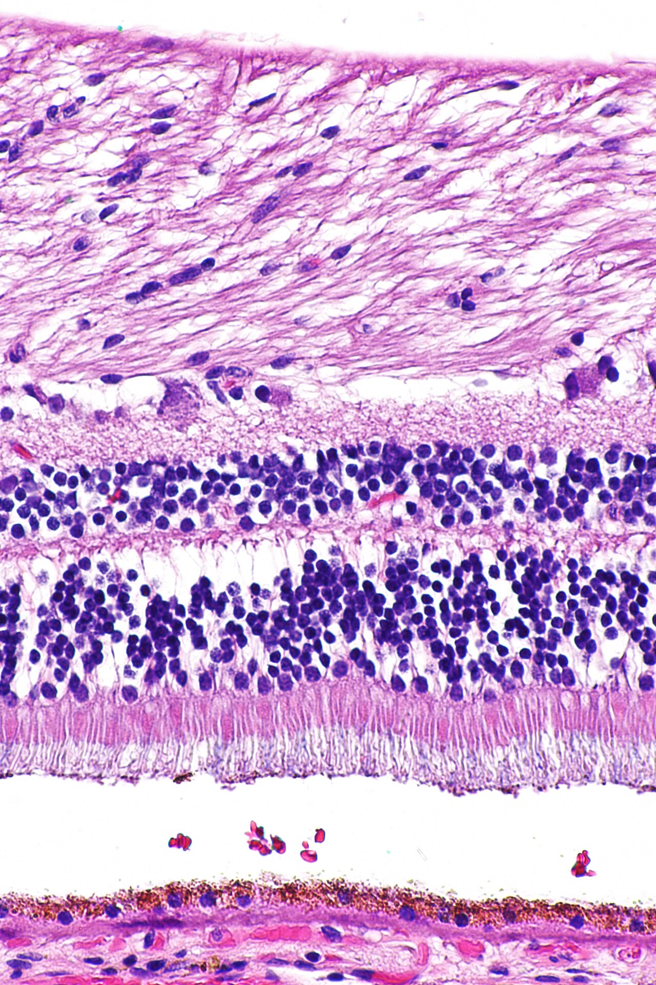
Image: Retina at high magnification, Librepath, CC BY-SA 3.0, via Wikimedia Commons.
Comments