

The limiting factor in synthetic tissue scaffolds is not stiffness, and it is not chemistry; it is the direction information travels. A hydrogel used to grow cells in three dimensions has its mechanics fixed before the cells arrive, and the cells must live with whatever was chosen. Real extracellular matrix does not behave that way. Cells rebuild it, and the rebuilt matrix feeds back and changes the cells.
A team working mainly at the Max Planck Institute for Polymer Research in Mainz has now made a material whose assembly is powered by the metabolism of the cells it surrounds. Their modified alginate stays liquid until it meets lactate, the waste product that heavily glycolytic tumour cells pour out, and then crosslinks into a covalent network precisely where that lactate is being produced. Around tumour spheroids the gel formed within a day, reached roughly a millimetre out as a measurable density gradient, held the spheroids at a constant diameter while controls kept growing, and shifted the cells' protein output from proliferation toward migration and matrix remodelling.
Why it matters: A material that assembles itself only where tissue is metabolically abnormal is, in principle, a way to act on diseased cells without having to target them molecularly. The nearest-term use is not treatment but honesty about models: tumour biologists currently study cells in scaffolds whose stiffness they chose themselves.
The study, by Braun et al. at the Max Planck Institute for Polymer Research, with collaborators at RWTH Aachen University and Johannes Gutenberg University in Mainz, appears in Nature Communications. It is an in vitro study throughout: cell monolayers, spheroids from two cell lines, and spheroids grown from resected human glioblastoma tissue.
Turning a metabolite into a chemical bond
The chemistry runs in two enzymatic steps. The researchers hung phenol groups onto sodium alginate by attaching tyramine, at about 2.5 mol% per sugar unit, low enough to keep the polymer soluble. Lactate oxidase converts lactate into pyruvate and hydrogen peroxide, and horseradish peroxidase spends that peroxide coupling neighbouring phenols into covalent junctions. Lactate is therefore not a signal the material listens for; it is the fuel it burns to build itself.
Driven with measured peroxide rather than cells, the response was sharp and bounded. Stiffness rose steeply between 0.5 and 1.5 mM peroxide, from 30 plus or minus 10 Pa to 4.8 plus or minus 0.5 kPa, a ceiling that matches the stoichiometric limit of pairing every available phenol. Omit either the enzyme or the peroxide and the modulus stayed near 10 Pa, indistinguishable from unmodified alginate. In real culture medium the same chemistry reached only 2.8 plus or minus 0.5 kPa, because medium tyrosine and serum proteins compete for the enzyme; numbers from a clean buffer would have overstated what happens around cells.
A gradient the cells write themselves
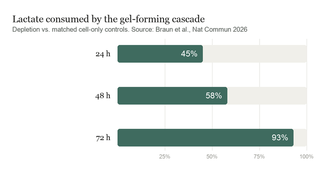
To separate gel from cells, the precursors were confined to a permeable insert while lung adenocarcinoma cells grew in the chamber below. Lactate in the shared medium was progressively consumed, and the insert's contents went from viscous liquid to self-supporting gel over three days, stiffening from 50 plus or minus 40 Pa to 2.9 plus or minus 1.3 kPa. Starve the cells of glucose and no gel formed, the cleanest available demonstration that metabolism, not the mere presence of cells, is the fuel line.
Around three-dimensional spheroids the geometry mattered more. Lactate does not dissipate evenly from a dense ball of cells, so gelation propagated outward as a graded network rather than a uniform block. Using tracer diffusion as a local probe, the team found a densely crosslinked zone within 100 to 400 micrometres of the surface, loosening out to about 1000 micrometres, where the gel ended. They call the implied concentration a lactate-equivalent rather than a true time-integrated measurement.
What confinement did to the cells
Encapsulated spheroids stopped expanding: controls grew from 474 to 507 micrometres over two days, while encapsulated ones went from 468 to 472 micrometres. But arrested growth is not the whole picture, and the paper does not pretend otherwise. ATP fell to 51 plus or minus 14% of controls, a resazurin readout to 75 plus or minus 15%, dead-cell staining rose substantially, and necrotic cores enlarged. Some of that size stabilisation is cells dying, not cells politely pausing.
The phenotype shift arrives with the control that makes it causal. Proteome profiling showed interleukin-8 strongly up, PAI-1 moderately up, and TIMP-1 and basic FGF down, a pattern consistent with migration and matrix remodelling rather than growth. Spheroid edges grew actin-rich protrusions, but only when the gel formed, and not in softer versions at 35 or 600 Pa, implying a stiffness threshold. Replacing the peroxidase with catalase, which destroys hydrogen peroxide before it can couple phenols, abolished both the gel and the proteomic changes. A second cell line gelled and shrank the same way but grew no protrusions, so the phenotype response is model-specific even where the material response is not.
What the study can't say yet
Nothing here happened in an animal. The patient-derived glioblastoma spheroids showed reduced invasive area over seven days, the most translationally suggestive result in the paper, but it is a small donor set in a dish. The authors' own list of gaps is long: no biodegradable linkers to remove the matrix afterwards, no data on where else in a body it might gel, no immune-competent model, and an unresolved oxygen problem, since lactate oxidase needs oxygen and the tumour regions richest in lactate tend to be hypoxic. The proteomics are relative abundances, so "more migration-associated protein" is not demonstrated invasion. On toxicity they are notably unwilling to oversell: Because prolonged exposure reduced metabolic activity and viable cell fraction, the system should not be described as intrinsically non-cytotoxic, the authors write.
Quick questions
Is this a cancer treatment? No. It is a laboratory material. The clinical version the authors sketch, a gel poured into a surgical cavity that stiffens where residual tumour cells are most glycolytic, is explicitly flagged as needing animal work first.
Why lactate rather than something more specific to cancer? Because it is abundant and it is fuel. The cascade needs chemical energy, not just a recognition event, and a metabolite produced in millimolar quantities can drive a reaction that a rare surface marker could not. Other oxidases would gate the gel on other metabolites.
One-line takeaway? Tumour cells' own metabolic waste can be made to build a covalent matrix around them, and that matrix then changes what the cells do, demonstrated convincingly in dishes and nowhere else yet.
Braun et al. "Cell-derived lactate triggers local hydrogelation and phenotype remodelling." Nature Communications, 2026. doi.org/10.1038/s41467-026-77702-x
PubMed PMID: 42805993.
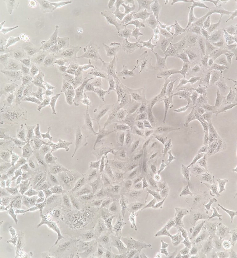
Image: A549 human lung adenocarcinoma cells in culture. Pinyapat Prisananuntakul, CC BY 4.0, via Wikimedia Commons.
Chart: lactate depletion data from Braun et al., Nature Communications 2026.
Comments