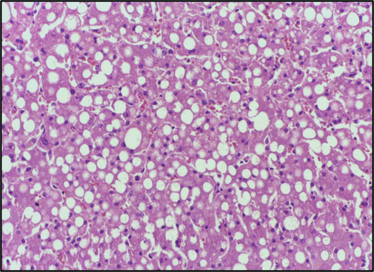
Fat that builds up in the liver is easy to picture and hard to treat. On a slide it looks almost decorative: hepatocytes ballooning with pale, round droplets, packed together like soap bubbles. What that picture hides is which genes are switched on, which cells are talking to each other, and where in the tissue the damage tips from harmless storage into scarring. A team led by researchers publishing in Nature Genetics set out to fill in that missing detail, one location at a time.
Metabolic dysfunction-associated steatotic liver disease, or MASLD, is now one of the most common chronic liver conditions on the planet. It runs on a spectrum. Some people simply accumulate fat, a state called MASL. Others progress to steatohepatitis, or MASH, where inflammation and scar tissue creep in. Predicting who moves along that path, and why, has been one of the frustrating open questions in the field.
Sixty-one livers, read cell by cell
The group profiled 61 human livers: 10 controls, 17 with MASL, and 34 with MASH. Rather than grinding the tissue into a slurry and measuring an average, they used single-cell and spatial transcriptomics to record gene activity while keeping track of exactly where each reading came from. On top of that they layered mass spectrometry imaging, which maps small molecules like lipids across the same slices of tissue. The result is a combined transcriptomic and metabolomic atlas, searchable online, that ties molecular signals to physical position inside the organ.
That spatial anchoring matters. A liver is not a uniform bag of cells. Blood arrives at the edges of each lobule and drains toward a central vein, and the chemistry shifts along the way. Averaging across the whole organ blurs exactly the local signals that drive disease.
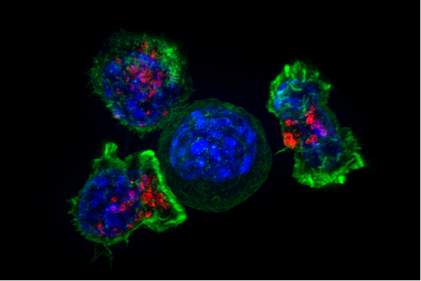
The macrophages that seem to help
The most striking finding concerns a population of immune cells the authors call lipid-associated macrophages, or LAMs. Macrophages in a diseased organ are often cast as troublemakers, stoking inflammation. Here the story runs the other way. The researchers identified a transcription factor, MITF, as a key regulator of how much fat these macrophages can take up and handle. And they found that the LAMs appear to protect the surrounding liver tissue, partly by secreting hepatocyte growth factor, a signal that supports liver cells.
So these cells are not just soaking up spilled fat. They seem to be actively defending the organ. That reframes them as a possible ally rather than a target to shut down, which is not how immune cells in a damaged liver usually get described.
The atlas also traced how scarring takes hold. By computationally pulling apart the spatial data, the team mapped a fibrosis-associated gene program that was concentrated in advanced MASH. Within the scarred zones, they saw signs of crosstalk between endothelial cells lining the central vein and hepatic stellate cells, the cells that lay down fibrous tissue. That points to a specific conversation between cell types that could push the liver toward irreversible damage.
On the metabolic side, the imaging showed that certain phospholipids pile up in a MASLD-specific pattern. The authors link that accumulation to phospholipid processing inside the same lipid-associated macrophages, tying the fat chemistry back to the immune cells at the center of the story.
What a map can and cannot do
An atlas is a description, not a verdict. This work identifies associations and cellular conversations in human tissue, but it does not prove that boosting LAM activity or blocking the stellate-endothelial crosstalk would change the course of disease in a living patient. The protective role of the macrophages rests on their gene expression and the growth factor they release; showing cause and effect will need experiments in models, not just snapshots of tissue. The samples also come from people already far enough along to have their livers biopsied, so the earliest steps of the disease are harder to capture here.
Still, the value of a resource like this is what other labs do with it. A searchable, spatially resolved map of human MASLD gives researchers a place to check where a gene of interest actually sits, which cells switch it on, and how that changes as fat gives way to scarring. For a disease with no shortage of failed drug candidates, knowing which cells to court and which to confront is a useful place to restart.
Comments