
Measuring metabolism in a living animal requires getting a labelled nutrient into its bloodstream and keeping it there for hours. Every established way of doing that changes the metabolism being measured. Continuous anaesthesia suppresses mitochondria. Surgical catheterisation of the jugular vein adds an operation, days of recovery and painkillers. Feeding the label produces uncontrolled intake. The authors state the problem without softening it: each of these approaches introduces physiological or technical variables that complicate accurate interpretation of in vivo flux.
A group at the Moffitt Cancer Center in Tampa did two things about it. They measured how much isoflurane anaesthesia actually distorts metabolism, across serum and four organs, and they built a delivery method that mostly avoids it: a tail vein catheter fitted in five to ten minutes under brief anaesthesia, which then runs for hours in an awake, freely moving mouse. Of 265 serum metabolites, 38 shifted at least twofold under two hours of isoflurane, 23 upward and 15 downward.
Why it matters: Stable isotope tracing is the standard method for measuring what metabolic pathways are actually doing, and results from it feed cancer and metabolic research broadly. If the anaesthetic used to collect the data reshapes the pathways, a large body of published flux measurements carries a distortion nobody quantified.
The work, by Kim et al., appears in Science Advances and used ten mice per group, five of each sex.
What the anaesthetic does
One pattern repeated across tissues. Acylcarnitines, which carry fatty acids into mitochondria for oxidation, fell consistently. Free amino acids rose, branched-chain amino acids among them. Pyruvate and tricarboxylic acid cycle intermediates fell. That combination is what mitochondrial inhibition looks like from the outside, and isoflurane is known to inhibit complex I of the respiratory chain.
The tissue-specific responses are more interesting than the shared one. The brain accumulated glycolytic and pentose phosphate intermediates while its acylcarnitines fell, which reads as a forced switch away from fat oxidation toward glucose. The liver looked busy detoxifying, with glucuronic acid up, and is where isoflurane is metabolised, but its amino acids, acylcarnitines and fatty acids were largely untouched. The pancreas showed the most extreme response, a near-global accumulation of free amino acids spanning almost every proteinogenic one.
The lung was the surprise. It receives the anaesthetic first and at the highest concentration, and it changed least. The authors flag it rather than bury it: Paradoxically, the lung showed the least metabolic perturbation of all tissues despite direct anesthetic exposure, suggesting that local exposure alone was not sufficient to drive broad metabolic remodeling in this tissue. Exposure and response are not the same thing, which is a useful corrective to the intuition that dose drives effect.
The tradeoff the method exposes
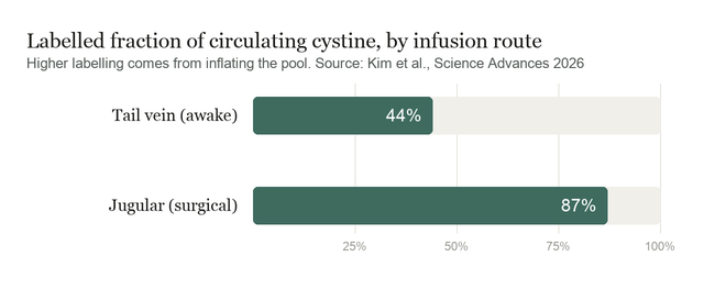
Comparing the new tail vein route against surgical jugular catheterisation produced a result that cuts both ways. Jugular infusion labelled circulating cystine far more heavily, close to 87% against roughly 44% for the tail vein. More label is better for detecting downstream products. But the reason the jugular number is higher is that the infusion inflated the circulating cystine pool itself, raising both labelled and unlabelled cystine well above baseline. The tail vein route kept the pool near physiological levels.
So the two desirable properties are in tension. Heavier labelling and an undisturbed metabolite pool cannot both be maximised, because the same flooding that drives enrichment is what moves the pool. A tracer experiment is supposed to follow metabolism without changing it, and the more confidently you can see the tracer, the more you have perturbed the thing you are watching. The paper demonstrates this clearly without quite framing it as the central finding.
Two controls make the new method credible. Brief isoflurane and catheter placement produced serum profiles that clustered with handled controls, while two hours of anaesthesia formed its own distinct cluster, so the short exposure needed for setup does not reproduce the damage. And running the same tail vein infusion in awake against anaesthetised mice left the isotopologue distributions largely comparable while pool sizes differed, which locates the anaesthetic's effect in metabolite abundance rather than in tracer incorporation.
What the study can't say yet
The method reduces perturbation rather than removing it. Catheter setup still requires brief anaesthesia, and serum corticosterone rose in both the tail vein and jugular groups compared with animals receiving no infusion at all, with no difference between routes. Being tethered to a pump for hours is itself a stressor, and this design cannot separate that from the route.
The tracing was demonstrated with one tracer, labelled cystine, followed into cysteine, glutathione, hypotaurine and taurine. Whether the route performs as well for glucose, glutamine or lactate, which are what most flux studies actually measure and which turn over far faster, is untested here. The lower enrichment may matter more for a rapidly cycling pool than for sulfur metabolites.
The anaesthesia survey is a single time point, two hours, in healthy young mice, with no recovery arm. How quickly the changes reverse is not addressed, which matters because many protocols anaesthetise only at the end. Four organs were profiled, so muscle, heart, fat and gut are absent, and metabolite abundance was measured rather than flux, meaning the study documents that pools moved without establishing which reactions changed rate.
The mechanism for the most striking result is explicitly unresolved. On the pancreatic amino acid accumulation the authors write that Whether the pancreatic pattern represents an amplification of a systemic response or a superimposed local mechanism warrants further investigation. Complex I inhibition is offered as a plausible contributor to the branched-chain amino acid rise, and the paper is clear that this was not directly tested.
Quick questions
What is stable isotope tracing? A nutrient made with heavier, non-radioactive atoms is infused, and mass spectrometry tracks where those atoms end up. It reveals which reactions are running, which measuring metabolite levels alone cannot.
Why does anaesthesia matter if both groups get it? In a comparison it partly cancels. The problem is describing normal physiology, or studying a tissue like brain or pancreas where the anaesthetic effect is large and may interact with whatever is being tested.
What's the one-line takeaway? Two hours of isoflurane shifted 38 of 265 serum metabolites at least twofold and reorganised brain and pancreas metabolism, and a tail vein catheter that works in awake mice avoids most of that, at the cost of labelling roughly half as much of the circulating pool.
Kim Y, Caldwell S, Long M, et al. "A simple, anesthesia-free infusion technique for in vivo metabolic tracing." Science Advances, 2026;12(37):eaeg1157. doi.org/10.1126/sciadv.aeg1157
PubMed PMID: 42726876.
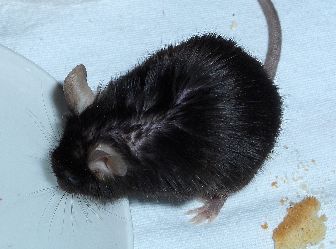
Image: laboratory mouse. Wualex, public domain, via Wikimedia Commons.
Comments