

The outside of a cell is coated in sugar. Chains of it hang off the proteins and lipids of the membrane, forming the densest chemistry on the cell's surface. Accounts of how the membrane is organised have mostly ignored this layer, treating structure as something proteins and lipids arrange between themselves while the sugars sit on top.
A collaboration across several Japanese institutes tested whether the sugars organise anything themselves, and found that they pair off with their own kind, and that the resulting pairs hold a well-known growth receptor in check.
Why it matters: If the outermost layer does structural work, it is a control mechanism sitting in the most accessible part of the cell, and one that has been missing from the standard picture.
Sugars that recognise themselves
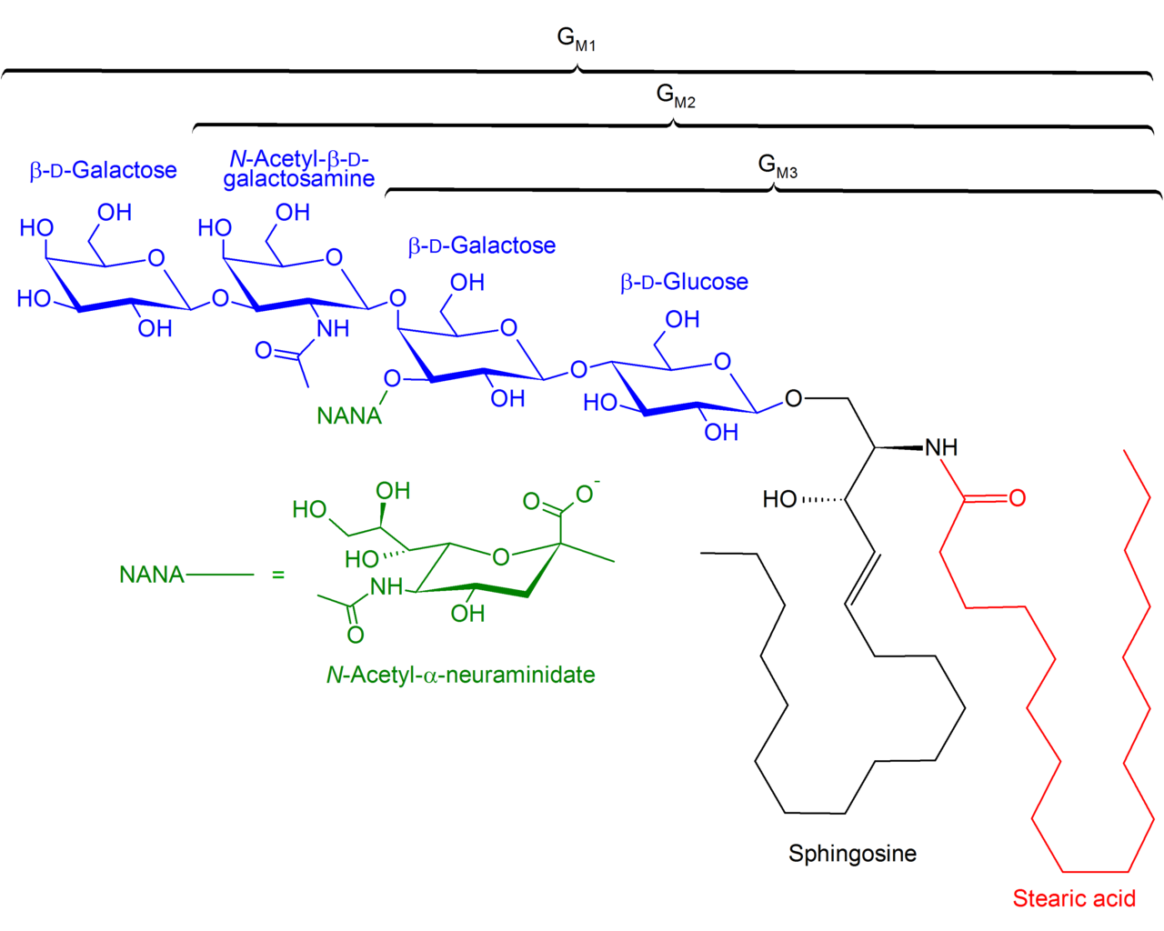
The problem with studying this has been chemical. You cannot label a natural sugar chain without changing it. The group instead synthesised 39 fluorescent versions of gangliosides, the sugar-bearing lipids of the membrane, and watched individual molecules move, both in living cells and in artificial bilayers where the composition is known exactly.
Every ganglioside they examined formed pairs with copies of itself. The pairing is brief and easily broken, but it is specific: each type prefers its own kind, and mixed pairs generally bound more weakly. Cholesterol stabilises these pairs into small clusters, which can go on to gather into groups of three and four.
There is a precedent worth noting. Certain membrane-tethered proteins were already known to form the same kind of fleeting pairs. But those are driven by proteins recognising proteins. Here the recognition happens between sugars, and because gangliosides and cholesterol are far more plentiful than those proteins, this is likely the larger contributor to the membrane's patchiness.
Holding a receptor in the off position
The functional half concerns EGFR, the receptor that drives cell growth and is mutated in many cancers. It works by pairing up; two receptors together switch on the signal.
The paired sugars bind to sugar chains at two specific positions on EGFR. The effect is to make receptor pairs form more slowly and come apart faster, which leaves more receptors as single units and suppresses the signalling that would otherwise happen without any growth factor present.
Two details make the mechanism convincing. Paired sugars bound the receptor roughly five times more readily than single ones, which is what you would expect if the point is having two binding surfaces rather than one. And the individual contacts are brief, lasting on the order of a tenth of a second, so the restraint comes not from any one durable bond but from a large number of fleeting ones in constant turnover.
The regulation also has an off switch built into it. After growth factor binds and the receptor pair locks into its stable form, the relevant sugar chains end up out of reach and the ganglioside can no longer pull the pair apart. It restrains idling and slows start-up, then stops interfering once the receptor is properly switched on.
One measurement gives useful context. At the receptor densities normally used in signalling experiments, the numbers indicate EGFR is mostly already paired even in resting cells. Something has to be keeping that population quiet, and this supplies a candidate.
The reach of this may be wide. GM3 is the dominant ganglioside of epithelial cells, which is also where EGFR is most broadly expressed, so the two are likely to sit together across much of the body. Several other growth receptors operate in the same sugar-rich context, which makes this a candidate general mechanism rather than a quirk of one receptor, though nothing here tests that directly.
What the study can't say yet
Why a sugar prefers its own kind is unexplained. The shared chemical groups are common to all gangliosides, so the selectivity must come from the three-dimensional shape and movement of the chains rather than their composition. As Suzuki and colleagues write in Nature Communications, Elucidating these features across diverse gangliosides remains an important subject for future investigation. The recognition is measured; its basis is not.
There is also a geometric puzzle the authors raise themselves. EGFR is largely excluded from the cholesterol-rich patches where these sugar pairs sit, so the two are not supposed to mix. The proposal is that the interaction happens at the boundaries between patches, which is plausible and untested.
One of the receptor's sugar attachment points is also a docking site for a protein that has the opposite effect, promoting clustering and activation. Whether the two compete, and what decides the outcome, is not addressed here.
And the work rests on synthetic analogs. They are close copies designed to behave like the real thing, but they are not the real thing, and results in engineered bilayers are cleaner than the crowded surface of a living cell for the same reason they are less representative of it.
Quick questions
What is a ganglioside? A lipid with a sugar chain attached, sitting in the outer half of the membrane with its sugars facing outward. They are abundant in most animal cells.
Why would slowing receptor pairing matter? Because pairing is what turns the receptor on. Anything that makes pairs form more slowly and break more readily raises the threshold for switching on by accident.
What's the one-line takeaway? Sugar chains on the cell surface recognise and pair with their own kind, forming small clusters that bind EGFR and keep it from activating without a signal, until the receptor locks into its active form and escapes.
Suzuki et al. "Cis glycan-glycan interactions organize membrane nanodomains that tune receptor signaling." Nature Communications, 2026;17(1). doi.org/10.1038/s41467-026-76857-x
PubMed PMID: 42660957.
Image: Structures of GM1, GM2 and GM3, LHcheM, CC BY-SA 3.0, via Wikimedia Commons.
Comments