
The limiting step in immunological memory is not the immune response, it is what survives the response. In an acute viral infection a handful of virus-specific CD8 T cells expand into a large population of killers, most of which dies off over the following weeks. The small surviving fraction is what makes a second encounter uneventful, and whether a given effector cell joins it is decided early. Diet is one candidate signal and the most loosely specified: the claim that fasting boosts immunity usually arrives with no mechanism attached.
A group at Seoul National University tried to pin one version of it down. Mice carrying transferred virus-specific T cells were infected with lymphocytic choriomeningitis virus, then cycled through 24 hours of complete food withdrawal alternating with 24 hours of free access to food, for the whole course of the infection. Thirty days later the fasted animals held more memory T cells, the increase concentrated in the central memory subset. The mechanism proved unexpectedly narrow: of everything fasting changes, the feature that mattered was reduced circulating glucose, and injecting glucose on the fasting days abolished the effect.
Why it matters: Engineered T cell therapies for cancer fail largely because the transferred cells run out of metabolic road inside a tumour. If a cheap nutrient condition during manufacturing imprints a longer-lived, exhaustion-resistant state, that is a lever on an expensive problem.
The study, by Park et al. at Seoul National University, appears in Science Advances. It was supported by Seoul National University, Korea's National Research Foundation and several government ministries, and the authors declare no competing interests.
Isolating glucose from everything else fasting does
Fasting changes many things at once, which is why dietary immunology is hard to interpret. The authors profiled plasma metabolites by nuclear magnetic resonance seven days after infection. The two groups separated clearly, but on one metabolite: glucose in the fasted group ran at approximately half the fed level, while citrate, beta-hydroxybutyrate, acetoacetate, acetate and alanine showed no differences between groups. Ketone bodies are the usual suspects in dietary restriction and here they did not move, which the authors attribute to alternating fasting and refeeding never establishing a sustained ketogenic state.
They then ran the test this field often skips: they put the glucose back. Injecting glucose on each fasting day restored plasma glucose to fed levels and abolished the whole phenotype, both the memory precursor proportion at day seven and the central memory pool at day 30. In culture the same lever worked directly. Naive T cells activated at 2 mM glucose rather than 8 mM were pushed toward memory precursors almost threefold, while KLRG1, the marker of terminal effector commitment, moved the other way.
Worse during the infection, better after it
The fasted animals were not mounting a larger response. At seven days they carried fewer virus-specific effector cells than fed controls in both spleen and lymph nodes, and those cells showed less Ki67, meaning they had divided less. Viral titres at three days did not differ significantly between groups, so fasting neither accelerated nor impaired control of the virus.
What changed was composition. Fasting skewed differentiation toward memory precursors, raised the IL-7 receptor alpha chain alongside CD122, which the authors note could alter responsiveness to the homeostatic cytokines IL-2, IL-7 and IL-15, and reduced both apoptotic and necrotic death during the contraction phase, when most of the effector pool is normally lost. On re-challenge, memory cells from fasted donors outnumbered fed-donor cells co-transferred into the same recipient and produced more interferon gamma and TNF alpha. Timing was strict: fasting during the effector window alone, or the contraction window alone, did not significantly improve memory at day 30.
A fuel switch written into chromatin
Effector cells from fasted mice carried less of the glucose transporter Glut1 and took up less glucose, carried more of the fatty acid transporter CD36 and took up more long-chain fatty acids, and accumulated lipid droplets at day seven that were largely depleted by day 30, consistent with stored lipid being burned later. Their oxygen consumption was higher, and in culture the equivalent advantage under low glucose was removed by blocking fatty acid oxidation with etomoxir.
The link to cell fate runs through histone acetylation. Low glucose raised H3K27ac, a mark of active regulatory regions, and that increase disappeared when fatty acid oxidation was blocked, placing the chromatin change downstream of the fuel switch. Accessibility shifted toward memory loci including Tcf7, Bcl6 and Eomes and away from glycolytic genes. Memory-like cells conditioned at low glucose and transferred into mice bearing ovalbumin-expressing melanoma were more abundant inside tumours, retained more TCF1, and included fewer cells positive for both PD-1 and Tim-3.
What the study can't say yet
This is a mouse study throughout, in female animals six to eight weeks old, and the protocol is total food withdrawal for a full day rather than any human eating schedule. The authors are explicit that their central manipulation is not clean, since moving blood glucose drags hormonal signalling with it: the relative contribution of glucose availability per se versus secondary systemic responses cannot be fully distinguished in the current study. Insulin and glucagon were not held constant.
The tumour arm is most open to over-reading. Those cells were conditioned in low-glucose culture, not harvested from fasting animals, so the experiment tests a nutrient condition as a manufacturing step, not fasting as a treatment. The target is ovalbumin in a melanoma line engineered to display it, a generous antigen compared with human tumour antigens, and the authors list validation in CAR T cells and clinical assessment as future work.
Quick questions
Should people fast to strengthen immune memory? Not on this evidence. The intervention was complete food withdrawal in mice, the readout a T cell population counted in spleen and lymph nodes, and no human was tested.
Why does the glucose injection experiment carry so much weight? It turns a correlation into a specific cause. Fasting alters many variables, but restoring one of them on fasting days erased the result, which rules out most of the others as the driver.
What is the one-line takeaway? Alternate-day fasting through a viral infection pushed mouse CD8 T cells toward long-lived central memory, the effect tracked an approximately halved blood glucose level and vanished when glucose was replaced.
Park et al. "Glucose restriction induced by intermittent fasting promotes alternative metabolic fluxes leading to memory CD8+ T cell development." Science Advances, 2026. doi.org/10.1126/sciadv.aeg0361
PubMed PMID: 42950055.
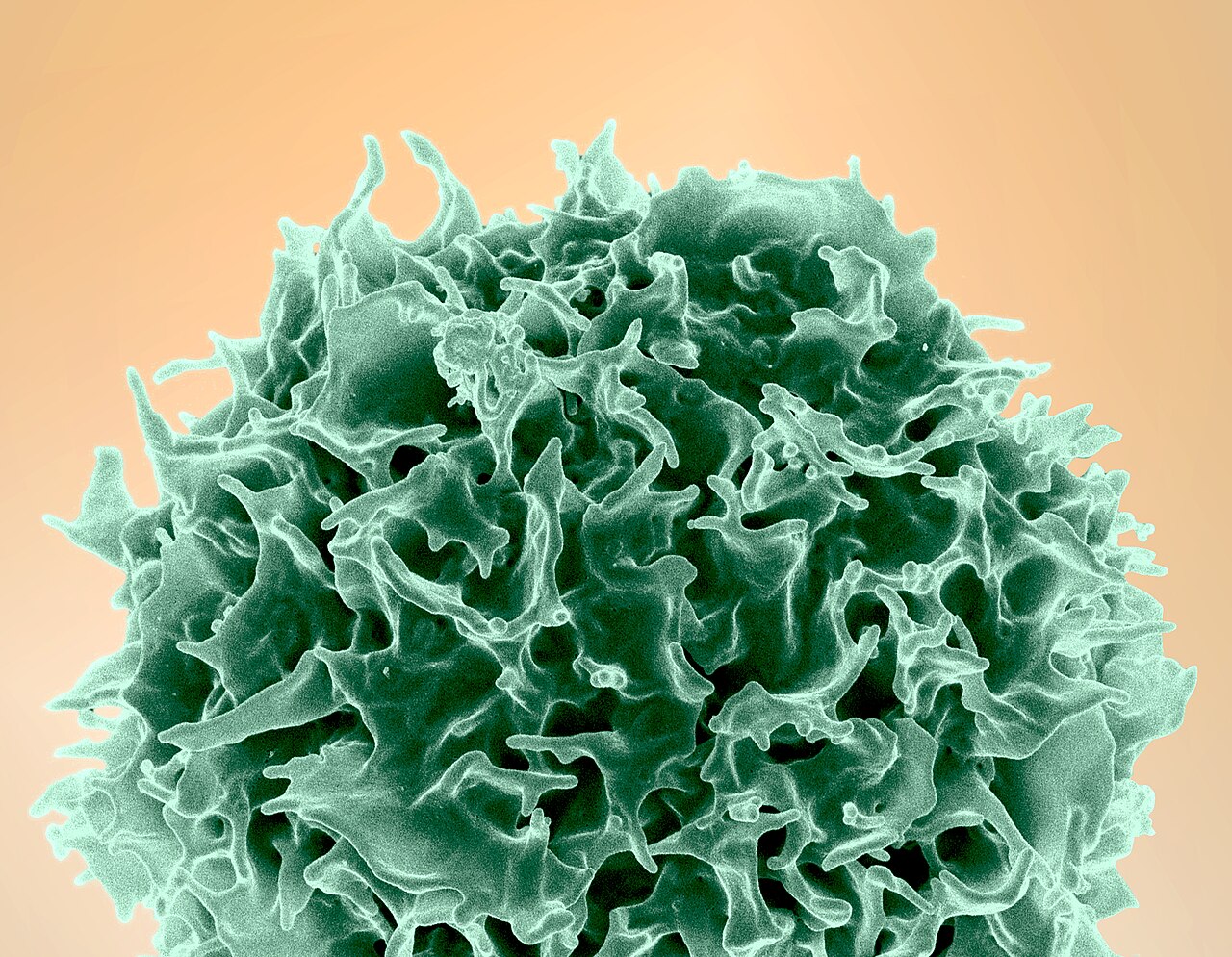
Image: scanning electron micrograph of a human T lymphocyte. NIAID/NIH, Public domain, via Wikimedia Commons.
Comments