

Contrast-enhanced MRI does not image a brain tumour. It images a leak. Gadolinium stays inside intact vessels, so the bright rim on a post-contrast scan marks where glioblastoma has broken the blood-brain barrier, not where its cells are. Glioblastoma's defining behaviour, though, is diffuse infiltration into brain that looks structurally intact. Extent of resection is one of the few modifiable predictors of survival here, and radiotherapy volumes are drawn around the same rim, so the gap between leak and cell is surgical rather than semantic.
A prospective single-centre study in Munich measured that gap by taking tissue. Sixty-one adults with IDH-wildtype glioblastoma had both contrast MRI and amino-acid PET before surgery, and 472 specimens were removed at navigated coordinates, matched back to both scans and graded by a neuropathologist blinded to the clinical data; the imaging was scored before any histology was merged in. Contrast MRI marked tumour in 52.6% of the samples that actually contained it. The PET tracer marked 91.2%. Of the 174 samples taken from tissue the MRI called unremarkable but the PET called positive, 171 held tumour.
Why it matters: Glioblastoma recurs at the edge of the resection cavity or along the border of the high-dose radiation field, the two lines drawn from the contrast scan. If viable tumour routinely sits outside those lines, the standard of care is aimed with an instrument that was never measuring tumour cells.
The study, by Stürzl et al. at the Department of Nuclear Medicine of LMU University Hospital in Munich, appears in Nature Communications; patients were enrolled between October 2019 and July 2025. Funding came from the German Research Foundation and the Thomas Kirch Stiftung, which the paper states had no role in design, analysis or reporting. Three authors declare competing interests, including lecture and advisory honoraria from pharmaceutical and imaging companies and, for one, research funding from Novocure and Telix Pharmaceuticals.
Two scans measuring two different things
The tracer is a fluorine-18 labelled analogue of the amino acid tyrosine, shortened to FET. Tumour cells import it through amino-acid transporters, and the signal was long attributed to one of them, LAT1. The authors say that is no longer the accepted account: current evidence treats it as a composite of several transporters plus tumour perfusion, microvascular architecture and barrier properties. A metabolic readout, but not a clean one.
Positivity was defined in advance, at a tumour-to-background ratio of 1.6 taken from the existing literature rather than fitted to this cohort. A threshold tuned on the data it is then evaluated on always flatters the test; re-derived from these samples, the optimal cut-off came out at 1.65, with an area under the ROC curve of 0.908. Uptake tracked histology in the expected order: maximum tumour-to-background ratios of 2.83 where both scans were positive, 2.22 where only the PET was, and 1.49 and 1.34 in PET-negative tissue.
Where the two scans disagree
Samples were sorted by what each scan said about the spot they came from: 210 from tissue both called positive, 174 from tissue only the PET called positive, 22 from enhancing tissue the PET called negative, and 66 from tissue neither flagged. In all 56 patients with any contrast enhancement, PET uptake extended past the enhancing border, and in 39 of those 56 it extended at least 10 mm beyond it. The positive predictive value in the non-enhancing PET-positive category was 98.3%, against 34.8% where both scans were negative.
Five patients had no contrast enhancement anywhere. All five were PET-positive, and all 30 of their PET-positive samples were tumour. The sharper comparison is with T2/FLAIR, the fallback map of infiltration that motivates resecting beyond the enhancing margin: among samples FLAIR-positive but neither enhancing nor PET-positive, 17 of 46 contained tumour, a positive predictive value of 37.0%. FLAIR marks oedema, gliosis and tumour alike.
The test is better at yes than at no
The gain is in detection, not in exclusion. The specificity of PET, 82.5% against 73.0% for contrast MRI, was numerically higher but not statistically distinguishable (p = 0.146), while the sensitivity gap was large and highly significant. Negative predictive value was 59.1% for PET and 19.2% for MRI, meaning that roughly four in ten PET-negative samples in this cohort still contained tumour. Detection also fell with cell density: PET found 99.1% of solid tumour samples but 82.3% of those from the infiltration zone, against 60.8% and 43.2% for contrast MRI, and uptake correlated only moderately with histological tumour burden (Spearman's rho 0.58). The authors put the inference plainly, suggesting that tumor below a cellularity threshold may fall beneath detection.
What the study can't say yet
Sampling was targeted rather than systematic, and deliberately so: extra biopsies of apparently inactive brain carry real surgical risk. PET-negative regions were therefore under-sampled, and specificity and negative predictive value rest on 88 such samples against 384 PET-positive ones. The authors also note that sampling was opportunistic rather than grid-based, with a median of five samples per patient and as many as 27. The diagnostic comparisons were computed sample by sample, so repeated samples from one person enter the tests as independent observations, which tends to understate the uncertainty. The recurrence subgroup was 16 patients and supplied no doubly-negative samples, so several comparisons could not be run in precisely the setting where treatment change is hardest to tell from tumour. MRI was acquired as routine clinical care at 1.5 or 3 tesla without a study protocol. And nothing here is an outcome: a randomised trial of PET-based against MRI-based targeting for re-irradiation of recurrent glioblastoma, cited by the authors, reported no advantage.
Quick questions
Should PET replace the MRI? No, and the paper does not argue it. MRI supplies the anatomy a surgeon navigates by, and PET proved unreliable at clearing tissue; the case is for reading both.
Would cutting to the PET margin help patients? Unknown. The study shows the extra tissue contains tumour, not that removing or irradiating it extends life; the one randomised test the authors cite showed no benefit.
What is the one-line takeaway? In 472 biopsies, the scan that defines glioblastoma surgery and radiotherapy found tumour in about half the samples that held it, while amino-acid PET found nine in ten and was almost always right when it saw tumour outside the enhancing margin.
Stürzl et al. "[18F]FET PET enables histopathology-validated detection of non-contrast-enhancing glioblastoma." Nature Communications, 2026. doi.org/10.1038/s41467-026-78980-1
PubMed PMID: 42995172.
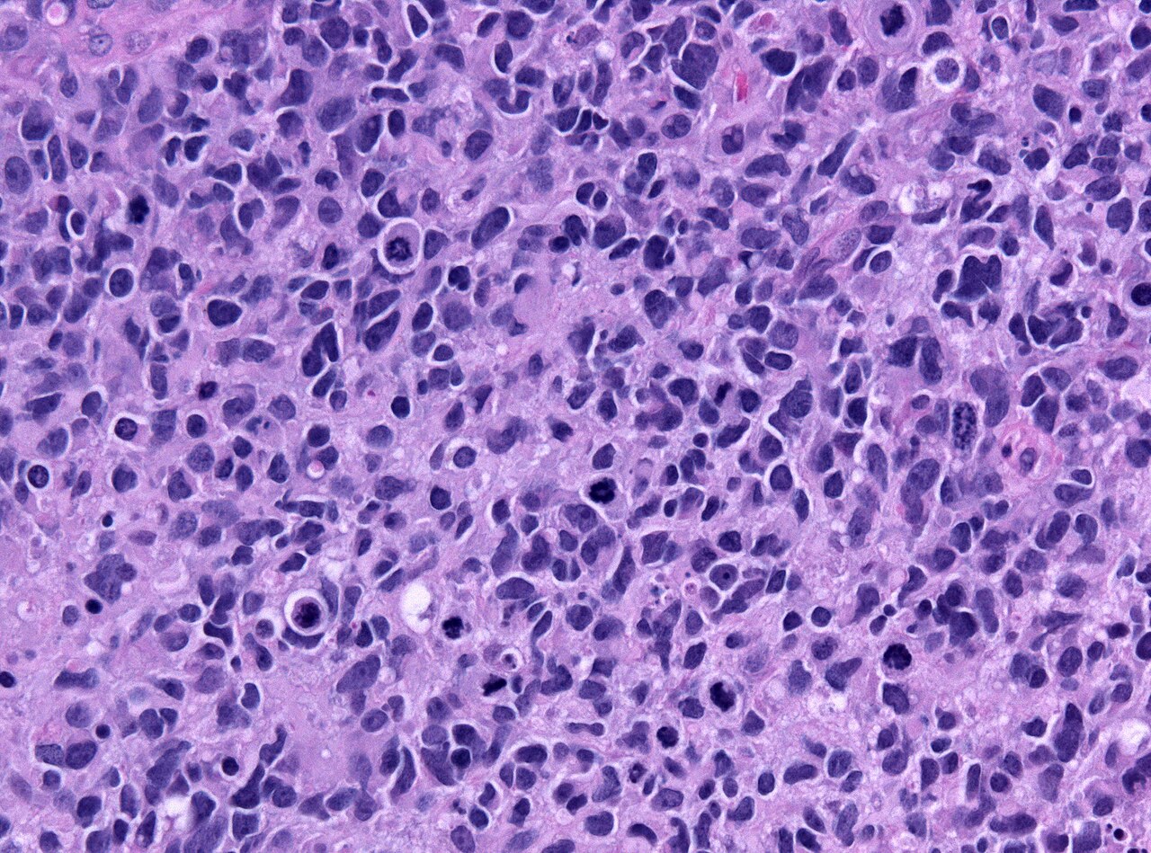
Image: glioblastoma histology. Jensflorian, CC BY-SA 3.0, via Wikimedia Commons.
Comments