A malaria parasite inside a red blood cell eats roughly 80 percent of its host's haemoglobin. That releases enormous quantities of free heme, which is toxic, so the parasite crystallizes it into an inert pigment called hemozoin. The process is essential, and it is what chloroquine and its relatives interfere with, which makes the question of how the parasite builds those crystals more than academic.
For about two decades the leading answer has been a protein called Heme Detoxification Protein, on the reasonable grounds that it converts heme into hemozoin-like crystals in a test tube. A group at the Bernhard Nocht Institute in Hamburg, working with collaborators in St Louis, Geneva and Mainz, has now tagged the protein at its own genetic locus and found it in the mitochondrion. Removing it does not affect hemozoin formation at all.
Why it matters: A protein believed to run an essential, druggable pathway turns out to run a different essential pathway. That changes what the parasite's heme chemistry is thought to depend on, and it is a clean case study in how a plausible in vitro result can outlive the evidence for it.
Why hemozoin is worth this much attention
The interest is entirely practical. Free heme generates oxidative damage and disrupts membranes, so a parasite consuming most of a red cell's haemoglobin has to neutralize an enormous chemical burden or die of it. Crystallizing heme into hemozoin is how it does this, and because the pathway is essential and has no human counterpart, it has been one of the most attractive drug targets in the parasite. Quinine, chloroquine and the artemisinins all owe some part of their action to interference with it.
That history explains why identifying a protein driver mattered so much. An enzyme is a target you can design against and assay against; spontaneous crystallization inside a lipid environment is far harder to work with. The search has produced three named candidates over thirty years, and all three now look like proteins that bind heme and do something else. The pattern is consistent enough to be a finding in itself.
What the protein actually does
Knocking it out produced a coherent set of defects, none of them about heme. Mitochondria depolarized. Parasites became hypersensitive to proguanil, an antimalarial whose activity depends on mitochondrial function. Development arrested. The decisive test was a rescue: supplying a route to pyrimidine synthesis that does not require the respiratory chain restored the parasites, which places the defect squarely in mitochondrial electron transport rather than anywhere near the digestive vacuole.
Electron flow was abolished outright, through loss of respiratory complexes III and IV. Those complexes contain subunits encoded by the mitochondrial genome and translated by mitochondrial ribosomes, so losing them selectively points at a translation failure rather than a respiratory one. Structural modelling combined with quantitative proteomics landed the protein inside the large subunit of the mitochondrial ribosome. It is a ribosomal protein that happens to bind heme, not a heme-processing enzyme.
How the misassignment survived so long
The paper is worth reading for its account of the evidence trail, which is more instructive than the correction itself. Test-tube activity came first: parasite lysates catalyze crystal formation, and this protein does so efficiently in isolation. The authors note drily that Although no clear rationale has ever been given for examining this protein in this context, detailed biochemical work followed, producing a model of exactly which histidine residues orient the heme monomers.
Localization then appeared to agree. Immunofluorescence and immunoelectron microscopy placed the protein in the host cytoplasm, in haemoglobin transport vesicles and in the digestive vacuole. A version expressed from a plasmid as a GFP fusion sat in a tubular compartment read as a haemoglobin trafficking intermediate. Co-immunoprecipitation found it associating with haemoglobin-degrading proteases. Four independent lines, all consistent, all pointing at the vacuole.
The difference here is endogenous tagging, which marks the protein at its native locus and leaves expression levels alone. Overexpressed from a plasmid, a protein can appear where the cell puts surplus copies rather than where it works, and antibody-based localization has its own failure modes in a parasite full of dense pigment. As Sarrazin and colleagues report in Nature Communications, tagging it properly moved it to the mitochondrion.
What the study can't say yet
The genetics were done in the rodent malaria parasite, which is the standard system for this kind of knockout work and is not the species that kills people. Mitochondrial biology is broadly conserved across Plasmodium, and the heme story is precisely the sort of thing that could differ between a parasite in a mouse and one in a human. Confirmation in the human parasite is the obvious next requirement.
The result is also more definite about what the protein does not do than about what makes hemozoin. Ruling out the leading candidate leaves the mechanism of crystal formation unresolved, and the two proteins previously proposed have already been ruled out on similar grounds. The field's current position is that an essential, drug-targeted process has no identified protein driver, which may mean it is largely spontaneous biochemistry rather than an enzyme-catalysed one.
Quick questions
Does this affect existing antimalarials? No. Chloroquine and related drugs act on the crystallization process itself, which still happens and is still essential. What changes is the explanation of how it is carried out.
How can a protein make hemozoin in a tube but not in a cell? Because heme crystallizes readily under the right conditions, and a protein that binds heme can promote it in vitro without doing so where it actually resides. Location is what the tube cannot tell you.
What's the one-line takeaway? Tagged at its own locus, the malaria protein long credited with building hemozoin turns out to sit in the mitochondrial ribosome, and deleting it leaves hemozoin untouched while collapsing respiration.
Sarrazin et al. "The Plasmodium heme detoxification protein functions in mitochondrial protein synthesis." Nature Communications, 2026;17(1). doi.org/10.1038/s41467-026-76511-6
PubMed PMID: 42567866.
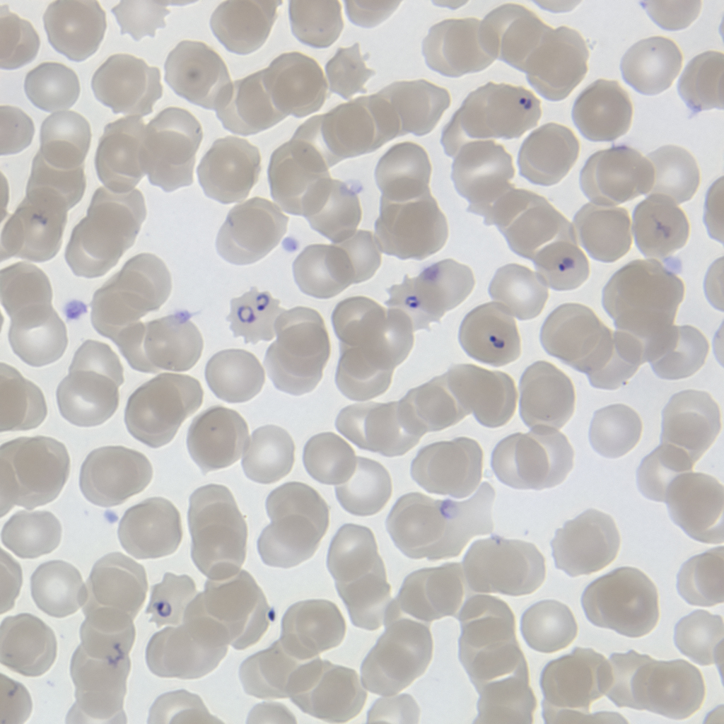
Image: Plasmodium falciparum in a blood smear, public domain, via Wikimedia Commons.
Comments