
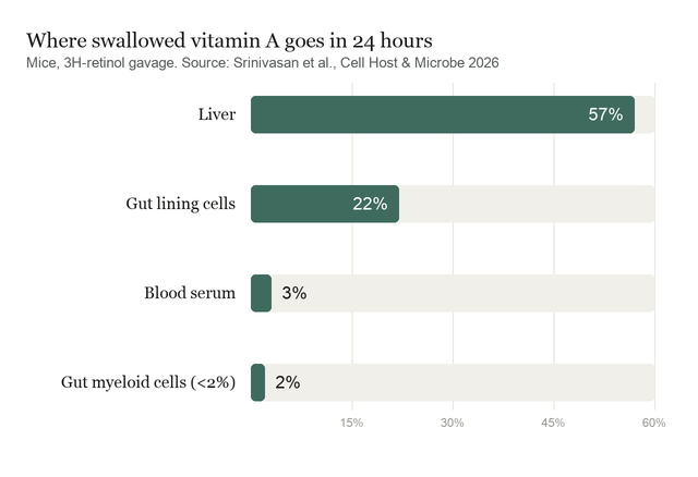
The limiting factor in vitamin A's effect on immunity is not how much an animal eats; it is how much arrives at the right cells. Vitamin A absorbed from food is claimed almost entirely by the liver, which stores it. In mice given a single dose of radiolabelled retinol, roughly 57 percent had reached the liver within 24 hours and about 3 percent was circulating in serum, while about 22 percent still sat in the gut lining and under 2 percent was detectable in the intestinal myeloid cells that pass the vitamin on to developing T cells. Against a liver share of 57 percent, that last figure is the whole immunological story.
A study published this month in Cell Host & Microbe shows that this sliver is not delivered passively but routed, and that the gut microbiota controls the routing. Working in mice, the authors traced retinoids along a three-step cellular relay running from the epithelial cells of the intestine, to CD11c+ myeloid cells in the lamina propria, to CD4+ T cells maturing in the mesenteric lymph nodes, a journey the paper reports is traversed over three days. Deplete the microbiota with antibiotics and absorption is unchanged while every downstream step of the handoff fails; restore the microbiota and the flux returns.
Why it matters: Vitamin A deficiency remains one of the world's most common causes of impaired immunity, and it has long been treated as an arithmetic problem of intake. This work argues that the delivery machinery matters independently of the supply, and that gut bacteria hold the switch.
The study comes from Srinivasan et al. at the University of Texas Southwestern Medical Center, with senior authors Lora Hooper, a Howard Hughes Medical Institute investigator, and Andrew Koh. It is a mouse study throughout, and its central tool is a radioactive tracer followed through sorted cell populations over ten days.
The control that localises the effect
Tracer studies are easy to over-read, since a signal can fall for many reasons. The design here is careful on that point. After gavage with tritiated retinol, radioactivity appeared sequentially in epithelial cells, then lamina propria myeloid cells, then myeloid cells in the mesenteric lymph nodes, then T cells there, and finally T cells back in the gut wall. Antibiotic treatment left uptake by the epithelium intact and left the serum and liver pools unchanged, but impaired movement through the myeloid–T cell axis. Because absorption and systemic distribution are held constant while only the local relay fails, the effect is pinned to the transfer steps rather than to how much vitamin A got into the animal. Reconventionalization, reintroducing a microbiota after antibiotics, rescued the movement, which rules out lasting antibiotic damage as the explanation. Liquid chromatography confirmed the depleted retinol content of myeloid cells directly, rather than relying on the tracer alone.
Two microbial signals doing two different jobs
The mechanistic core of the paper is a separation that most accounts of microbial immune education blur. The first signal is generic. Microbe-associated molecular patterns, the conserved bacterial structures the innate immune system detects, induce epithelial cells to express serum amyloid A proteins, which bind retinol and deliver it to myeloid cells through the receptor LRP1. Mice lacking all SAA isoforms, and mice lacking LRP1 on CD11c+ cells, both showed reduced retinoid accumulation in myeloid cells while epithelial uptake stayed normal, establishing necessity. For sufficiency, transgenic mice expressing Saa1 in the epithelium regardless of colonisation restored retinoid loading and downstream gene expression even under antibiotics. SAA, in other words, is the microbiota's instruction, and the instruction works without the bacteria present.
The second signal is specific, and this is the more interesting result. SAA loads myeloid cells and sends them to the lymph nodes, but it does not complete the delivery. Passing retinoids to T cells required microbial antigen, and required cell contact. The pathway is thus gated twice: an innate phase that positions the courier, and an antigen-dependent phase that authorises the handoff to a particular T cell. The vitamin is therefore not broadcast to the immune system; it is issued to T cells being activated by microbial material at that moment.
Why the timing points to early life
The pathway is not constant. Saa1 expression rose between two and six weeks of age in conventional mice, the window spanning weaning and microbial expansion, while genes for other retinol-binding proteins stayed flat or declined. Retinoid accumulation, receptor activity in lymph-node T cells, and CD4+ T cell numbers in the small intestine all rose over the same period, and every one of those increases was absent in SAA-deficient and germ-free animals. The reading the authors offer is that this system exists to synchronise immune maturation with microbial colonisation rather than to run continuously.
What the study can't say yet
Every experiment here is in mice, including the knockouts and transgenics that carry the causal weight, and nothing in the paper demonstrates the pathway in humans. The authors are also explicit that the precise chemical form of vitamin A handed to T cells was not directly measured; the model that myeloid cells convert retinol to retinoic acid before transfer rests on converging indirect evidence, since retinoic acid degrades too quickly in light to quantify reliably in isolated cells. The mechanism of transfer itself is unresolved: contact and antigen are required, and the immunological synapse is proposed as the site, but that remains a hypothesis. One author declares consulting and funding ties to nutrition and pharmaceutical companies, disclosed in the paper and not attached to any product claim in it.
On the division of labour, the authors state the limit of their own first signal plainly: "SAA alone is insufficient to mediate retinoid transfer to T cells; antigen recognition is required for this final step."
Quick questions
Does this mean vitamin A supplements will not work without the right gut bacteria? No. The experiments manipulate the delivery pathway in mice, not supplementation outcomes in people, and intake was not the variable tested.
Why would the body make delivery this complicated? The two-step gate ties a scarce nutrient signal to evidence of real microbial exposure, so T cells receive it as they respond rather than at random.
What is the one-line takeaway? In mice, gut bacteria control whether dietary vitamin A reaches maturing intestinal T cells, using one signal to load the courier and a second to authorise delivery.
Srinivasan T, Dende C, Ruhn KA, et al. "The gut microbiota directs vitamin A flux to regulate intestinal T cell development." Cell Host & Microbe, 2026;34(8):1630-1645.e9. doi.org/10.1016/j.chom.2026.05.019
PubMed PMID: 42309057.
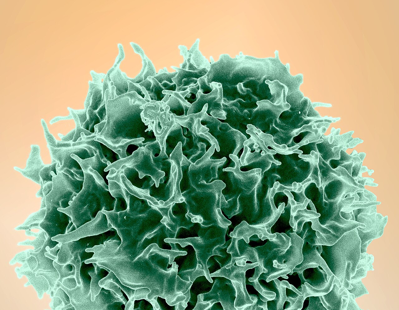
Image: colorized scanning electron micrograph of a human T lymphocyte. NIAID, CC BY 2.0, via Wikimedia Commons.
Comments