
Almost all the effort in gene editing has gone into the editor. Cas9 was made more precise, then base editors removed the need to cut at all, then prime editors widened what could be written. Meanwhile the step that actually limits most therapies has had far less attention: getting the machinery inside the cell, out of the compartment it lands in, and into the nucleus. A lipid nanoparticle that binds a cell but never releases its cargo has delivered nothing.
A group at Wisconsin-Madison went looking for what the cell does to obstruct that. They built a genome-wide screen linking the disruption of 19,114 genes directly to whether an edit happened, and found six genes whose removal makes editing markedly better. Deleting the top candidates raised efficiency roughly sixfold.
Why it matters: If the limiting factor is the cell's own machinery rather than the editor's chemistry, then dose, toxicity and cost in gene therapy are all governed by targets nobody has been optimizing. These are not better editors; they are the locks the existing ones are being pushed against.
Screening for delivery rather than repair
Earlier screens of this kind mostly profiled DNA repair, because they used viral delivery or reporters already integrated into the genome, which quietly skips every physical barrier. This one was built the other way round, so uptake, trafficking and nuclear import stayed part of what was being measured. The readout is deliberately blunt: was there an indel or not, for each of 19,114 gene perturbations.
Bluntness is a virtue here. A screen that scores editing outcomes in detail invites arguments about which repair pathway produced which product. A screen that asks only whether an edit occurred cannot be biased in that way, and what came back was a coherent group of membrane and trafficking proteins rather than the repair factors previous work had surfaced.
The control that makes the claim
A gene whose loss improves editing could be acting on delivery or on anything downstream, including repair itself. The team separated those with an experiment worth describing, because it is the load-bearing part of the paper. They repeated the comparison using electroporation, which forces the editor through the membrane directly and skips endocytic uptake entirely.
If these genes were affecting repair, the knockouts would still edit better under electroporation. They did not. For every top candidate but one, knockout and normal cells performed the same once the membrane was bypassed, which locates the effect at entry. As Saxena and colleagues put it in Nature Communications, the electroporation data argue against a dominant contribution from altered DNA repair kinetics or intrinsic Cas9 activity under these conditions, although such effects cannot be definitively excluded. The hedge is appropriate and the design is the right one.
The identity of the strongest hit is unexpected. GJB2 encodes connexin 26, a gap junction protein, which is not where anyone would look for a brake on nanoparticle delivery. The second, BET1L, is a vesicle trafficking protein, which fits the story more comfortably. Imaging supported the mechanism: more Cas9 had accumulated inside cells six hours after delivery when the gene was absent.
Tested where it would matter
The demonstrations move deliberately from convenient to clinically relevant. In cell lines, depleting either gene improved base editing sixfold, both when correcting a pathogenic mutation and when installing one, showing the effect is about getting the editor in rather than about any particular edit.
The harder test used patient-derived cells modelling a retinal channelopathy, edited with lipid nanoparticles, the delivery vehicle actually used in the clinic. Knocking down either gene improved efficiency more than 3.5-fold, and in a subset of edited cells the Kir7.1 ion channel worked again. That is a smaller gain than in cell lines, which is the expected direction and worth stating plainly.
What the study can't say yet
The strategy has an obvious problem the paper does not solve: these genes were removed by knockout or knockdown before editing. Deleting a gap junction protein from a patient's retina to make a therapy work better is not a treatment, it is a second genetic intervention with its own consequences, and connexin 26 mutations already cause deafness. Turning this into something usable requires transient inhibition, and nothing here demonstrates that.
Restoration of channel function also occurred in a subset of edited cells, not throughout the tissue, and the study reports no functional outcome beyond the cellular level. Whether a 3.5-fold delivery gain translates into clinical benefit depends on where the therapeutic threshold sits for a given disease, which varies enormously and is not addressed. The screen was also run in one cellular context; the authors validated across payloads and editors, which is more than most, but not across the full range of tissues a therapy would target.
Quick questions
Why does removing a gene make editing work better? Because some cellular proteins impede the uptake or intracellular routing of the delivery particle. Removing them lets more editor reach the nucleus.
Could this be used in patients? Not as done here. The genes were knocked out beforehand, and a therapy would need to inhibit them temporarily and reversibly, which this work does not demonstrate.
What's the one-line takeaway? Screening 19,114 genes for what blocks non-viral editing found six cellular brakes on delivery rather than repair, and removing the top two raised editing efficiency about sixfold in cell lines and over 3.5-fold with lipid nanoparticles in patient-derived cells.
Saxena et al. "Genome-wide CRISPR screening identifies cellular factors controlling nonviral genome editing efficiency." Nature Communications, 2026;17(1). doi.org/10.1038/s41467-026-76350-5
PubMed PMID: 42595755.
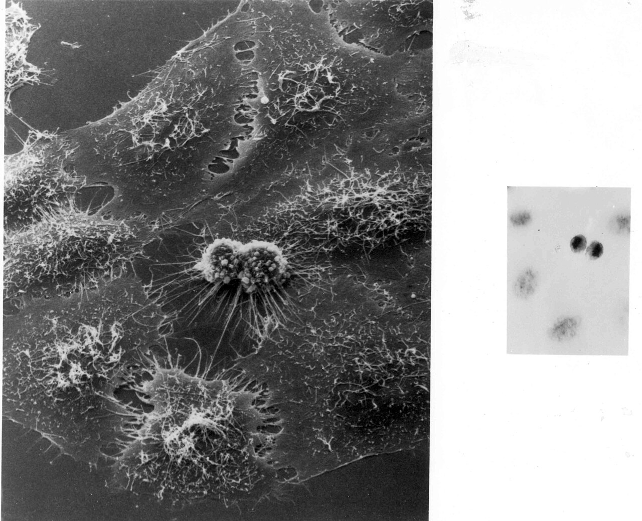
Image: HeLa cells, National Institutes of Health, public domain, via Wikimedia Commons.
Comments