
Most of what an animal does with light leaves no trace. Behaviour does not fossilise, eyes rot, and the sensory world of something that died three hundred million years ago is usually beyond reach. There is one exception worth exploiting: melanosomes, the small packets of melanin that pigment tissue and shield it from light, are tough enough to survive in rock. If their shape tracks what they are for, then a fossil melanosome is a fragment of preserved physiology.
A group at University College Cork has tested that premise properly, by measuring melanosome geometry across living fishes first and only then turning to fossils. Digesting tissue from a range of species, they found that melanosomes from internal organs keep much the same shape across every major group examined, which they read as a functional constraint that has held for a very long time. The eye was the exception. In bony fishes the eye melanosomes are cylindrical; in jawless and cartilaginous fishes they are only ever spheroidal.
Why it matters: This is a method as much as a result. If melanosome shape reliably indicates tissue and lineage, palaeontologists gain a way to read physiology and light environment off fossils rather than inferring them from skeletal anatomy, which is largely silent on the subject.
The study, by Cirtina et al. at University College Cork, appears in Nature Communications. It combines measurements from extant species with fossil specimens from the late Carboniferous Mazon Creek deposits.
Shape as a clue to function
The cylindrical shape in bony fishes is associated with retinomotor movement, the repositioning of pigment and photoreceptors as light levels change, along with shielding photoreceptors and sharpening acuity. The spheroidal-only pattern in jawless and cartilaginous fishes fits a different strategy, favouring sensitivity and regulating light by other means, including adjusting the pupil. Two solutions to the same problem, distinguishable by the shape of an organelle.
One organism stood out. The authors report that Larger, more abundant melanosomes in lungfish likely reflect adaptations that underpin the vertebrate water-to-land transition, lungfish being the living group closest to the lineage that moved onto land. Moving from water to air means a brighter, harsher light environment and more ultraviolet, so more pigment is the expected direction, and the hedge in the word likely is appropriate: this is one group's measurements lining up with a known evolutionary event.
What the study can't say yet
The clean two-category picture is cleaner in summary than in the data. On the comparisons between tissues and taxa, the authors are explicit that the distributions sit on top of each other, indicating overlapping morphological distributions and suggesting that differences reflect shifts in the central tendency rather than clear morphological separation. A difference in averages across overlapping distributions is a real signal and a poor basis for classifying an individual fossil melanosome.
Their statistics make the same point in a stricter way. Attempting to resolve the finer geometric differences into distinct groups failed: Mixture modelling using the ICL criterion did not resolve these subtle geometric differences into discrete populations, and thus they were treated collectively here. The categories are analytical conveniences at that level of detail rather than things the data separates on its own.
Then there is the date. The fossils are Carboniferous, roughly three hundred million years old, and the headline claim reaches back to the Ordovician around 445 million years ago. That is not a measurement but an inference: the lineage differences are present in the Carboniferous material, so they must have arisen before those lineages diverged, which was much earlier. The reasoning is sound and the conclusion is still a projection well beyond the oldest specimen in hand.
The fossil record itself is the remaining constraint, and the authors name it plainly: Limited sampling and preservation bias in the available fossil material, however, cannot be ruled out. Mazon Creek preserves soft tissue unusually well, which is why it was used, and that same unusualness makes it a poor sample of Carboniferous life in general. Whatever conditions favoured preservation there may also have favoured particular kinds of animal.
Quick questions
Can you really tell how an extinct fish saw from a fossil? Partly, and indirectly. What the shape of a preserved melanosome supports is an inference about which tissue it came from and which lineage, and from there about light-handling strategy. It is not a reconstruction of vision.
Why would melanin be involved in seeing at all? Because managing stray light is half of seeing. Pigment behind and around the photoreceptors absorbs light that would otherwise scatter, and moving that pigment is one way eyes adapt between bright and dim conditions.
What is the one-line takeaway? Melanosome shape is conserved in the internal tissues of fishes but splits by lineage in the eye, a split visible in Carboniferous fossils and argued to date to the Ordovician, on distributions that overlap substantially and a fossil sample the authors agree may be biased.
Cirtina et al. "Melanosome evolution shaped sensory biology and terrestrial adaptations in early aquatic vertebrates." Nature Communications, 2026. doi.org/10.1038/s41467-026-78877-z
PubMed PMID: 42946737. Full text: PMC13714305.
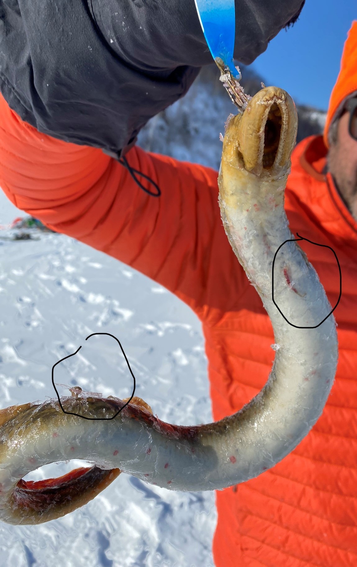
Image: sea lamprey (Petromyzon marinus). Rob Foster, CC BY 4.0, via Wikimedia Commons.
Comments