Make a piece of tissue transparent and you can, in principle, look straight through it. Tissue clearing does exactly that. Soak a mouse brain or a tumor biopsy in the right chemistry and it turns glassy, letting light travel deep instead of scattering off in the first fraction of a millimeter. The catch has always been the microscope. To image a cleared block the size of a sugar cube in three dimensions, you need a machine that stays sharp in every direction, moves fast, and does not choke on the optical distortions that clearing solutions introduce. Most setups pick one or two of those. A team at the University of Gottingen has now built one that manages all three, and did it largely with parts you can order from a catalog.
The paper appeared in Nature Biotechnology on November 13, 2025, from the lab of Jan Huisken, one of the people who helped popularize light-sheet imaging in the first place. Light-sheet microscopy works by illuminating a sample with a thin plane of light from the side while a camera watches from above, so only the slice in focus gets lit. It is gentle and quick. But when you want fine detail across a large cleared sample, the light sheet has to be swept along the optical axis, and that sweep tends to be slow and to blur as it goes.
What the numbers actually say
The new microscope reaches 850-nanometer resolution that is isotropic, meaning it is the same whether you measure across the sample or down through it. That evenness matters. Many instruments look crisp in one plane and smeared in the perpendicular one, which quietly distorts anything you try to trace or measure in 3D. It handles samples up to a cubic centimeter and copes with refractive indices from 1.33 to 1.56, the range that different clearing recipes land in, so you are not locked into one protocol.
Two engineering choices carry most of the weight. To keep the image sharp across a wide field, the team put a concave mirror in the remote-focusing part of the system to cancel out the curvature of the light sheet, which roughly doubled the usable field of view. To make it fast, they drove the light sheet's motion with closed-loop feedback rather than an open sweep. That pushed the speed up tenfold, to 100 frames per second, without giving back resolution or field of view. The optical trick underneath is an axially swept light sheet paired with an air objective and a simple meniscus lens, a combination that delivers diffraction-limited performance and corrects aberrations at the same time.
Why off-the-shelf is the point
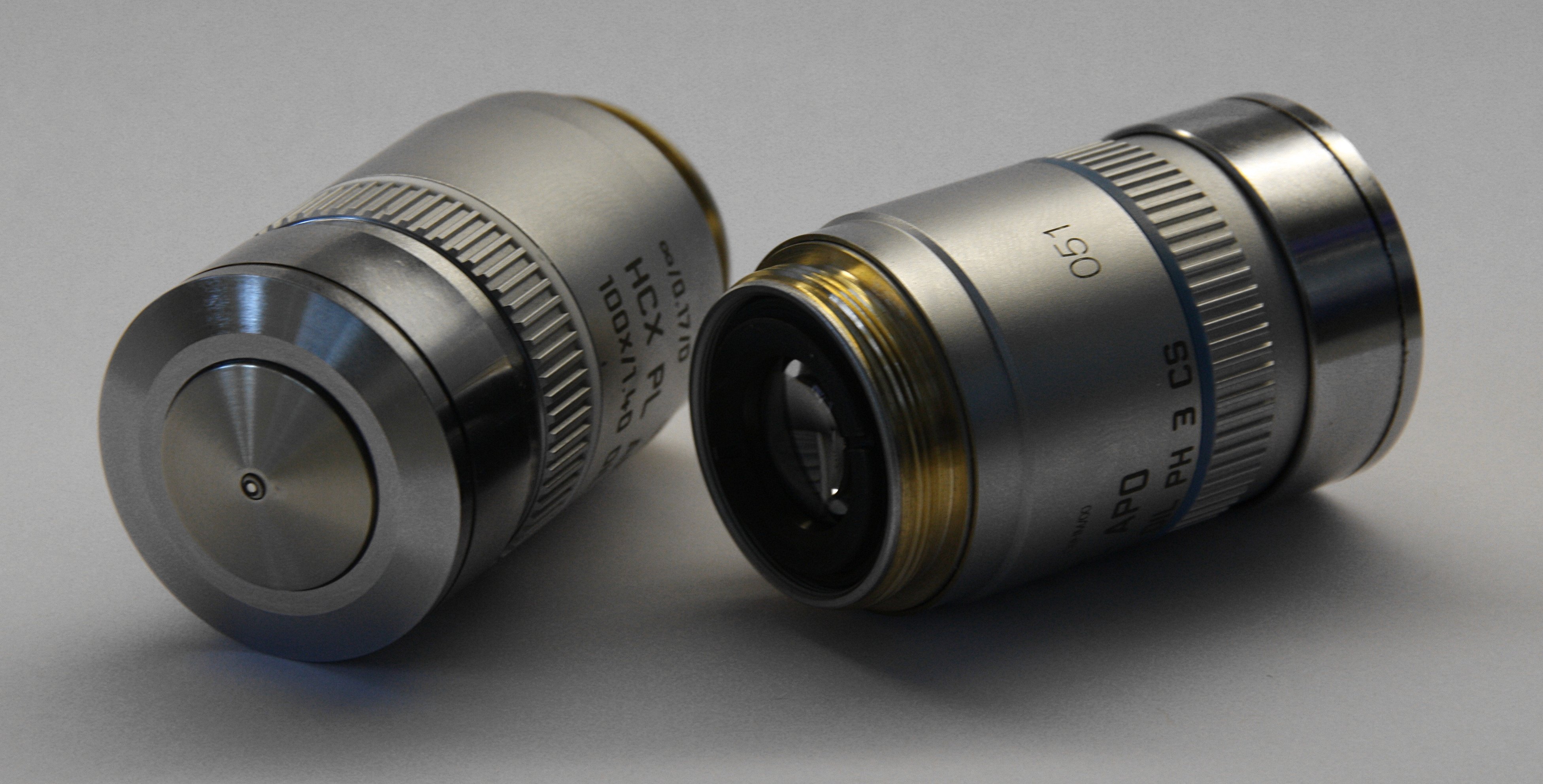
Specialized microscopes often depend on custom-ground optics that cost a fortune and cannot be replaced when they break. Here the authors leaned on standard components. An air objective. A meniscus lens. A concave mirror. That decision is not just thrift. It means other labs can plausibly build or adapt something similar without a machine shop and a grant line item for bespoke glass. The team benchmarked the instrument across a wide span of scales, from subcellular structures up to the full centimeter, using several different clearing methods, which is the kind of stress test that separates a demo from a tool people can actually adopt.
Fast, even, deep imaging of cleared tissue is useful well beyond pretty pictures. It is how you map the fine branching of blood vessels through an intact organ, follow neural wiring across a whole brain region, or check how a drug or disease has reshaped a piece of tissue in full three dimensions rather than in thin slices reconstructed by hand.
The honest caveats
A few things are worth keeping in mind. This is an instrument paper. The demonstrations show the microscope resolving structure across scales in cleared, fixed samples, not a validated pipeline for any particular clinical or biological question. Eight hundred fifty nanometers is a real improvement in isotropy for centimeter-scale work, but it is not the tens-of-nanometers regime of super-resolution methods, so the smallest molecular details remain out of reach. Clearing itself can shrink or swell tissue depending on the method, which any quantitative measurement has to account for. And a microscope that produces cubic-centimeter volumes at 100 frames per second generates enormous datasets, so the bottleneck often shifts downstream to storage and analysis. None of that undercuts the result. It just marks where the work goes next.
Comments