


The hard constraint on a multi-cancer blood test is not whether it can see cancer. It is the specificity you are willing to demand, because that single choice silently fixes everything else. Set the false-positive rate where a screening programme needs it and sensitivity falls; relax it and every negative result carries less weight while the follow-up burden rises. A headline sensitivity figure means nothing until you know the threshold it was read at.
GutSeer, developed across five Chinese medical centres and reported by a group at Zhongshan Hospital, Fudan University, is a blood test for five gastrointestinal cancers: colorectal, oesophageal, gastric, liver and pancreatic. It reads 1,656 methylation markers covering 33,032 CpG sites, and extracts a second class of signal from the same sequencing run, the physical fragmentation pattern of circulating DNA. In a validation cohort it reached 82.8% sensitivity at 95.8% specificity. The number that matters more appears further down: raise specificity above 99%, the level suited to population screening, and sensitivity drops to 64.0%.
Why it matters: Gastrointestinal cancers account for roughly a quarter of cancer cases and a third of cancer deaths, and are usually found late. A blood test that triages who needs an endoscopy would be useful even if it never becomes a screening test, but those are different claims and rest on different numbers.
The study, by Huang et al., appears in Molecular Cancer and enrolled 3,386 participants: 1,355 with gastrointestinal cancer, 63 with advanced precancerous lesions and 1,900 without cancer. Early-stage disease was well represented, 64.0% of cancers at stage I or II, which matters because that is where a blood test earns its place and where circulating tumour DNA is scarcest.
One test, or five with different competence
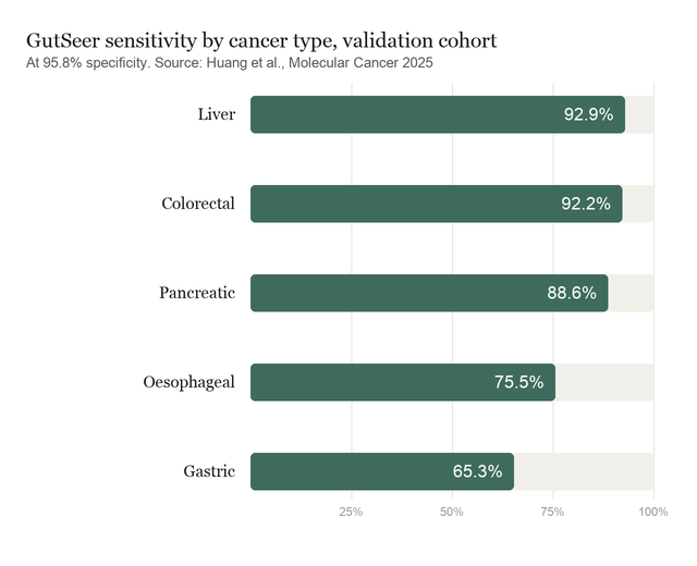
The aggregate sensitivity conceals a wide spread. In validation, GutSeer detected 92.9% of liver cancers and 92.2% of colorectal cancers, but 75.5% of oesophageal and 65.3% of gastric. A blind test on 846 prospectively collected samples reproduced the pattern almost exactly, at 91.3%, 74.1% and 65.2%. A third of gastric cancers were missed twice over, in two independent cohorts, which makes it a property of the assay rather than a sampling accident.
Restricting to early disease widens the gap further. Stage I and II sensitivity in validation ran from 54.4% for gastric cancer to 90.4% for liver. Against advanced precancerous lesions, the stage where detection would change the most, the test found 8 of 17 colorectal cases, 7 of 18 oesophageal and 6 of 28 gastric. Those denominators are small enough that the percentages should be read as directions, not estimates.
Tissue-of-origin prediction, which decides where the follow-up endoscopy points, was 82.4% accurate overall but 64.1% for pancreatic cancer. Locating the tumour is a second problem, and the test is meaningfully worse at it for the cancer with the worst prognosis.
What the comparison does and does not establish
Three results give the approach real support. First, combining methylation with fragmentation beat either alone, with an area under the curve of 0.958 against 0.929 and 0.934, and the difference held under a formal test. Second, against a published whole-genome fragmentomics method on the same samples, GutSeer scored 0.963 to 0.887, and 79.6% against 59.0% on tissue of origin, from a panel small enough to run cheaply. Third, the test beat the serum markers still used in clinics, CEA, CA19-9 and AFP, in every comparison.
The authors are clear about the design choice underlying the headline. a slightly lower specificity (e.g., 95%) could be more practical for targeted cancer detection in outpatient settings. That is a defensible position for a triage tool used on people who have already presented with symptoms. It is not the specificity a screening test applied to healthy people would need, and the paper's own 99% figure shows what that costs.
What the study can't say yet
The cohort is not a screening population. Participants were recruited from inpatient and outpatient care at five hospitals, so they had already entered the medical system. Sensitivity and specificity measured there describe discrimination among people who presented, not the predictive value of a positive result in an asymptomatic population, where cancer is far rarer and false positives dominate.
Cancer and control groups also differed in ways that predict cancer by themselves. In the test cohort, cancer patients were older, 62.2 against 55.0 years, and more often male, 63.8% against 49.5%, both differences significant. The authors' own multivariate analysis found age and sex to be independent predictors. This is handled rather than ignored: propensity matching on age and sex held the area under the curve at 0.938, which is the right check and largely answers the objection. But it is an adjustment, not a design that avoided the imbalance.
Total DNA quantity and unique molecule counts were also significant predictors, which is expected since cancer raises circulating DNA, and they carried less predictive power than the model. Bisulfite conversion imposes DNA input requirements and may itself bias the fragmentation measurements the test partly depends on. Per-cancer cohorts remain small despite the overall size. And the controls were never followed forward, so a control who develops cancer next year is counted today as a false positive. The authors list that last one plainly: a longitudinal follow-up of tested healthy controls is essential to reduce misclassification.
Quick questions
What is fragmentomics? Circulating DNA is cut into fragments, and the lengths and end sequences differ between tumour and normal tissue. Reading that pattern alongside methylation extracts two signals from one sequencing run.
Why does the specificity threshold matter so much? Because almost everyone screened is healthy. At 95% specificity, one in twenty healthy people tests positive, and in a low-prevalence population those outnumber the true cases. Tightening the threshold cuts false positives but, here, costs about 19 points of sensitivity.
What's the one-line takeaway? A compact methylation panel detected 82.8% of five gastrointestinal cancers at 95.8% specificity in a hospital cohort, but only 64.0% at the specificity a screening programme would require, and only 65.3% of gastric cancers at either.
Huang A, Guo DZ, Su ZX, et al. "GUIDE: a prospective cohort study for blood-based early detection of gastrointestinal cancers using targeted DNA methylation and fragmentomics sequencing." Molecular Cancer, 2025;24(1):163. doi.org/10.1186/s12943-025-02367-x
PubMed PMID: 40468355. ClinicalTrials.gov: NCT05431621.
Image: DNA double helix. Via Wikimedia Commons.
Comments