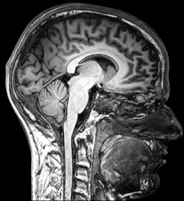
Diffuse midline glioma is one of the cruellest diagnoses in pediatric medicine. The tumours grow in the pons and thalamus, deep in the developing brain, and almost every child dies. Surgeons usually cannot cut them out. What has been missing is a way to tell, early on, which children face the fastest course and why.
A team led by Jai Sidpra and colleagues, reporting in Nature, offers an unusual answer. They treated the tumour not as a lump to be measured but as something plugged into the brain's wiring. Their method, called tumour network mapping, uses standard brain imaging to work out which healthy circuits a given glioma is connected to. The result is a shared pattern they call the DMG network, and how tightly a tumour couples to it tracks with how long a child survives.
Cancer that taps into living circuits
The starting point comes from lab work over the past decade showing that these gliomas are not passive. In animal models, they form real connections with neurons through synapses, and they link to each other through gap junctions. Electrical and chemical signals from active brain circuits then feed the cancer, driving it to grow and spread. The tumour, in effect, wires itself into the brain and lets normal activity do some of its work.
Whether that picture holds in living children, and whether it matters at the bedside, was open. Sidpra and colleagues mapped the brain-wide connectivity of pontine and thalamic gliomas and found a conserved network across both tumour locations. Tumours sitting in high-connectivity positions were tied to shorter survival. When the researchers tested the measure in two separate patient groups they had not used to build the model, the tumour's functional connectivity to the DMG network still predicted overall survival on its own.
The developmental timing lined up too. The places in the brain where in-network metabolic activity peaks during childhood matched the ages when these tumours most often appear. Looking at single-nucleus RNA sequencing, the more connected tumours were enriched for genes involved in synapses, the physical junctions the cancer seems to exploit.
A clue hiding in surgical accidents
One finding stands out for how concrete it is. When surgeons happened to remove some high-connectivity thalamic tumour tissue during a procedure, those children did meaningfully better. That is a striking hint. If the most electrically integrated parts of the tumour are the ones fueling its growth, then knowing where they are could tell surgeons and radiation planners where to aim. Right now, treatment for these tumours has very little to steer by.
The appeal here is that the input is a brain scan, not a fresh biopsy. Sampling a tumour buried in the brainstem is dangerous, and many children never have their tumour tissue removed at all. A prognostic read built from imaging that many patients already get is a different kind of tool. It could help sort children by risk, guide who might benefit from more aggressive local treatment, and give families and doctors a clearer sense of the road ahead.
What the map can and cannot do yet
This is a prognostic marker, not a treatment. Knowing a tumour is highly connected does not by itself make it easier to remove or destroy, and the surgical-resection observation came from incidental cases rather than a planned trial. That signal needs to be tested directly before anyone changes how these operations are done. The work also rests on connectivity models and existing scans across several cohorts, so how it performs when built into routine clinical workflows, on different scanners and in different hospitals, still has to be shown.
There is a bigger idea underneath the numbers. If a childhood cancer grows by borrowing the brain's own circuits, then the circuits themselves become part of the disease, and part of what we can measure. Reading a tumour by the company it keeps, rather than by its size alone, points toward treatments aimed at cutting those connections. For a disease that has resisted almost everything, a map of where the tumour has plugged itself in is a place to start.
Comments