

Cells dispose of damaged parts by wrapping them in a membrane and digesting them, a process called autophagy. A small protein named LC3 does the sorting. It gets anchored into the growing membrane by a lipid tail, and receptors carrying the cargo dock onto it through a short sequence that slots into two hydrophobic pockets on its surface.
Those pockets have been described in detail by structural biology, but almost always with LC3 floating free in solution. In the cell it is not free. It is stuck to a membrane, and a group at CSIR-IGIB in New Delhi asked whether being stuck there changes it.
Why it matters: If a protein only adopts its working shape once anchored, then structures solved in solution describe a version that never does the job, and the regulation happens somewhere nobody has been looking.
A site that appears on contact
The group ran atomic simulations of LC3 in three conditions: free in solution, anchored to a membrane, and anchored with a cargo receptor bound. The membrane was built to resemble a real cellular one rather than a generic bilayer, mixing four lipid types in the proportions found in the endoplasmic reticulum. Each condition was run in triplicate for a microsecond, nine microseconds in total, with further runs using two other cargo receptors.
Anchoring reorganised the protein globally. Contact with lipids rearranged the electrical interactions along its membrane-facing surface, which shifted contacts throughout the structure and opened the two pockets that receive cargo. In solution those pockets are partly closed.
Tracing how the change propagated led to a specific region on the far side of the protein, a loop-and-strand stretch that is floppy in solution and becomes ordered when the protein sits on a membrane. Once ordered, its conformation controls the distant binding pockets. That is allostery: two sites, physically separated, communicating through the body of the protein.
The mechanism has an unusual feature. Lipids do not plug into this site. They pass near it. As Gahlot and colleagues report in Nature Communications, dynamic and transient lipid engagement in the vicinity of an allosteric site is sufficient to induce its ordering. Fleeting, non-specific contact is enough.
Testing it by breaking it
A simulation showing correlated motion is suggestive, not conclusive. The correlation could reflect something else changing both regions at once.
So they engineered the site. Using the simulations to guide design, they built variants with three residue substitutions each, one set intended to destabilise the site and one to stabilise it, and both were designed to leave membrane anchoring intact so that only the regulatory region differed.
Both worked as intended. The stabilised variant bound cargo receptors better and sequestered more cargo, confirmed by super-resolution microscopy, electron microscopy and assays of autophagy activity. The destabilised variant sat on the membrane and did essentially nothing.
The crystal structure of the active variant closed the loop. It showed the regulatory site locked in the predicted arrangement and, importantly, small rearrangements in the distant binding pocket, which is what the simulation said should follow. The prediction and the structure agree on both ends of the connection.
The design approach is worth noting too. Rather than screening many mutants and keeping whichever worked, the team used the simulated ensemble of shapes to pick residues predicted to lock the site into one state or the other, then tested that specific prediction. When both variants behaved as designed, the model earned more credit than a single confirming experiment would have given it.
Why it is interesting beyond autophagy
Lipid-controlled allostery is familiar in ion channels and receptors, proteins built into membranes with lipids pressing against them from all sides. LC3 is a small globular protein tethered to a membrane by a tail. The finding that it works the same way suggests the mechanism is not restricted to proteins embedded in membranes, and that peripheral proteins may routinely be doing something in their anchored state that solution structures miss.
What the study can't say yet
The mechanism was discovered in simulation, and simulations depend on their assumptions. The force fields, the lipid composition and the microsecond timescale all constrain what can appear. The crystal structure and the functional assays anchor the conclusion at both ends, but the intermediate steps, the actual path by which the signal travels, come from computation and have not been observed directly.
What triggers ordering is also not pinned down. Saying that transient lipid contact near the site is sufficient describes a condition, not the specific interactions responsible, and no particular lipid has been identified as necessary. Whether cells modulate this by altering membrane composition is an obvious question the study does not address.
The engineered variants are also tools rather than states the cell uses. They demonstrate that the site controls binding, which is what they were built to test. Whether anything in a living cell tunes this site, and what would do the tuning, is unknown.
Quick questions
What is allostery? When binding or change at one part of a protein alters behaviour at a distant part, through the protein's own structure rather than direct contact.
Why does anchoring to a membrane matter? Attachment holds the protein against a surface and changes which parts of it are exposed and how they move. Here that is enough to open the sites cargo receptors use.
What's the one-line takeaway? LC3 has a regulatory site that only takes shape once the protein is on a membrane, and engineering that site alone switches cargo capture on or off without disturbing the anchoring.
Gahlot et al. "A programmable lipid-triggered allosteric site modulates LC3 LIR receptor binding activity." Nature Communications, 2026;17(1). doi.org/10.1038/s41467-026-76697-9
PubMed PMID: 42665581.
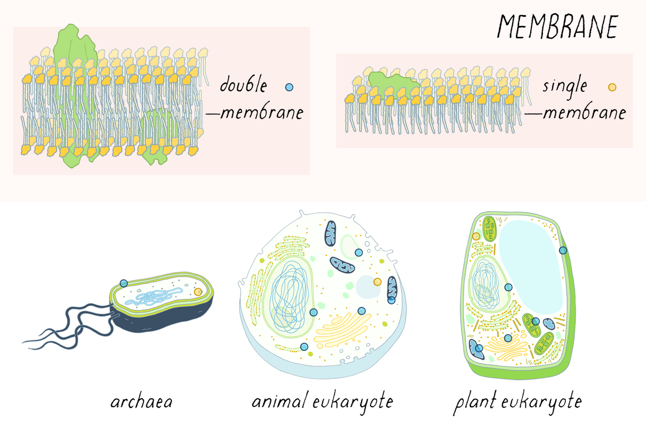
Image: Lipid bilayer membrane illustration, explorebiology, CC BY 4.0, via Wikimedia Commons.
Comments